File:Amniote rhodopsin 3D structures.png
Jump to navigation
Jump to search
Size of this preview: 555 × 600 pixels. Other resolutions: 222 × 240 pixels | 444 × 480 pixels | 711 × 768 pixels | 1,077 × 1,164 pixels.
Original file (1,077 × 1,164 pixels, file size: 1.15 MB, MIME type: image/png)
Captions
Captions
Add a one-line explanation of what this file represents
Summary
[edit]
| Description |
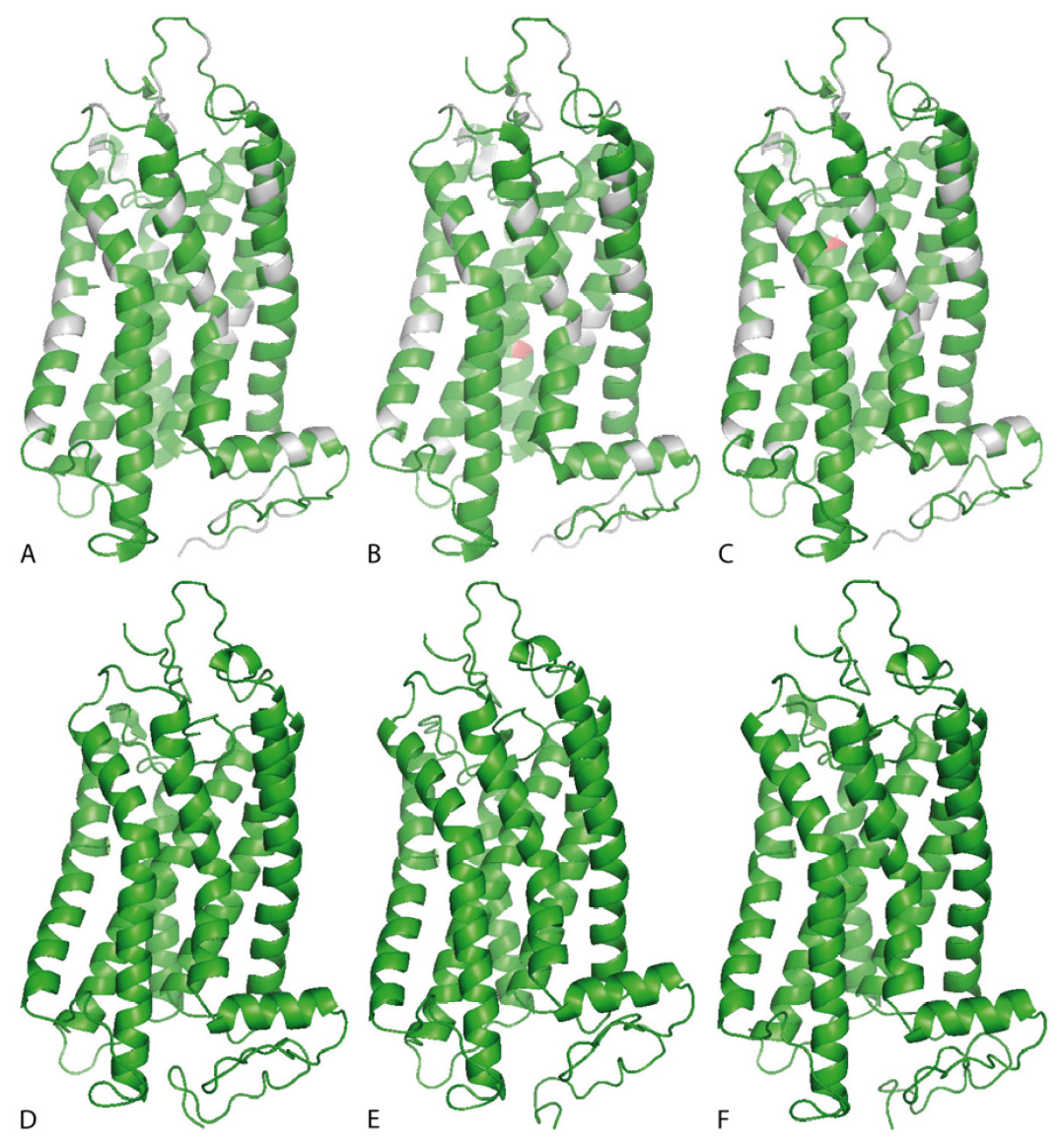
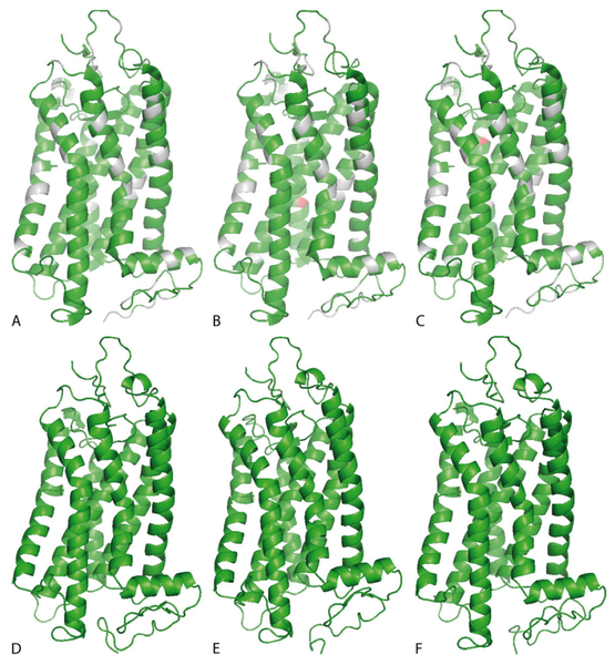
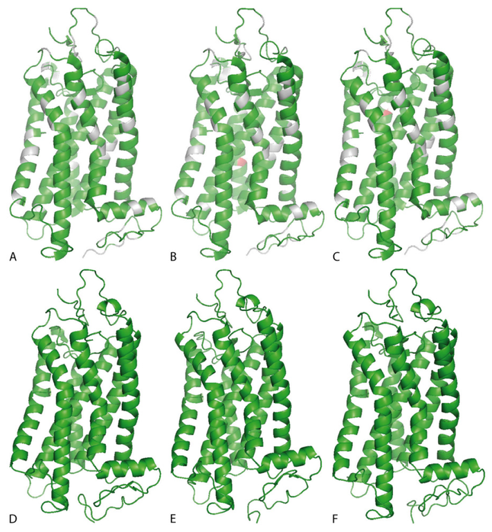
English: Original figure caption: Rhodopsin 3D structure of all pigments from this study [visualized by means of PyMOL Molecular Graphics System, V 1.3]. (A) shows the echidna rhodopsin with amino acids differing from bovine rhodopsin highlighted in gray. Red marks indicate the substitutions of mutants (B) T158A and (C) F169A. (D-F) Ancestral pigments, i.e. (D) Amniota, (E) Mammalia, and (F) Theria.
Deutsch: 3D-Struktur verschiedener realer und rekonstruierter Rhodopsin-Moleküle, erstellt mit Hilfe von PyMOL Molecular Graphics System, V 1.3. (A) Rhodopsin des Schnabeligels (Tachyglossus aculeatus). Aminosäuren, die von denen des Rhodopsins des Hausrindes abweichen, sind grau hervorgehoben. Rote Markierungen zeigen Substitutionen bei den Mutanten T158A (B) und F169A (C). (D-F) Anzestrales Rhodopsin des letzten gemeinsamen Vorfahren der Amniota (D), der Mammalia (E) und der Theria (F). |
| Date | |
| Source | Fig. 21 in: Visual pigment evolution and the paleobiology of early mammals. PhD thesis, Naturwissenschaftliche Fakultät der Humboldt-Universität zu Berlin, 2011 (PDF 4.5 MB) |
| Author | Constanze Bickelmann |
| Permission (Reusing this file) |
The source document of this image is published on the edoc server of the Humboldt-Universität Berlin under a CC-BY-SA-3.0 license |
Licensing
[edit]
This file is licensed under the Creative Commons Attribution-Share Alike 3.0 Unported license.
- You are free:
- to share – to copy, distribute and transmit the work
- to remix – to adapt the work
- Under the following conditions:
- attribution – You must give appropriate credit, provide a link to the license, and indicate if changes were made. You may do so in any reasonable manner, but not in any way that suggests the licensor endorses you or your use.
- share alike – If you remix, transform, or build upon the material, you must distribute your contributions under the same or compatible license as the original.
File history
Click on a date/time to view the file as it appeared at that time.
| Date/Time | Thumbnail | Dimensions | User | Comment | |
|---|---|---|---|---|---|
| current | 19:59, 8 August 2016 | | 1,077 × 1,164 (1.15 MB) | Gretarsson (talk | contribs) | {{Information |Description ={{en|1=Original figure caption: ''Rhodopsin 3D structure of all pigments from this study ''[visualized by means of PyMOL Molecular Graphics System, V 1.3.]''. (A) shows the echidna rhodopsin with amino acids differin... |
You cannot overwrite this file.
File usage on Commons
There are no pages that use this file.
File usage on other wikis
The following other wikis use this file:
- Usage on eu.wikipedia.org