File:Anaplastic astrocytoma.jpg
Jump to navigation
Jump to search
Size of this preview: 800 × 552 pixels. Other resolutions: 320 × 221 pixels | 640 × 442 pixels | 1,024 × 707 pixels | 1,200 × 828 pixels.
Original file (1,200 × 828 pixels, file size: 200 KB, MIME type: image/jpeg)
Captions
Captions
Add a one-line explanation of what this file represents
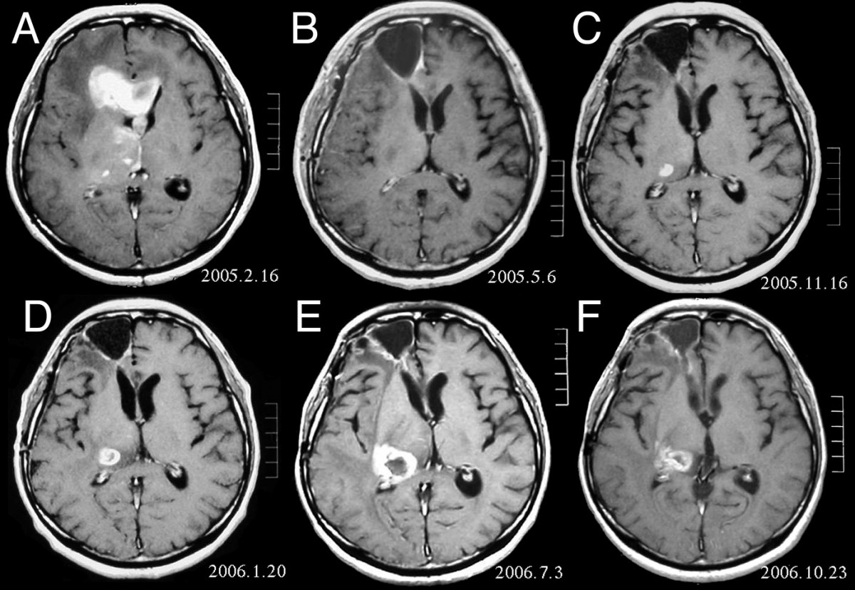
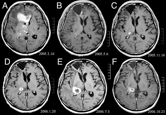
| Description | MRI of brain. (A) Initial MRI on February 16, 2005, shows a tumor in the right and left frontal lobe as well as the right thalamus. (B) MRI after surgery, radiation and chemotherapy. The tumor has completely disappeared except for slight enhancement adjacent to the surgical margin. (C) Recurrence of the thalamic tumor despite maintenance chemotherapy on November 16, 2005. (D) Increase in size of the thalamic tumor two months after stereotactic radiotherapy. (E) After 6 cycles of TMZ therapy, the thalamic lesion enlarged, and the patient developed dysarthria and hemiparesis. (F) After 2 courses of treatment with interferon-beta and TMZ, the tumor shows a partial response. |
| Date | |
| Source | Fujimaki T, Ishii H, Matsuno A, Arai H, Nakagomi T.Effectiveness of interferon-beta and temozolomide combination therapy against temozolomide-refractory recurrent anaplastic astrocytoma.World J Surg Oncol. 2007 Aug 4;5:89. PMID 17683572 doi:10.1186/1477-7819-5-89 |
| Author | Fujimaki T, Ishii H, Matsuno A, Arai H, Nakagomi T. |
| Permission (Reusing this file) |
BioMedCentral License |
This file is licensed under the Creative Commons Attribution 2.0 Generic license.
- You are free:
- to share – to copy, distribute and transmit the work
- to remix – to adapt the work
- Under the following conditions:
- attribution – You must give appropriate credit, provide a link to the license, and indicate if changes were made. You may do so in any reasonable manner, but not in any way that suggests the licensor endorses you or your use.
File history
Click on a date/time to view the file as it appeared at that time.
| Date/Time | Thumbnail | Dimensions | User | Comment | |
|---|---|---|---|---|---|
| current | 16:47, 25 February 2008 | | 1,200 × 828 (200 KB) | Filip em (talk | contribs) | {{Information |Description=MRI of brain. (A) Initial MRI on February 16, 2005, shows a tumor in the right and left frontal lobe as well as the right thalamus. (B) MRI after surgery, radiation and chemotherapy. The tumor has completely disappeared except f |
You cannot overwrite this file.
File usage
The following page uses this file:
Global file usage
The following other wikis use this file:
- Usage on ar.wikipedia.org
- Usage on bg.wikipedia.org
- Usage on cs.wikipedia.org
- Usage on da.wikipedia.org
- Usage on de.wikipedia.org
- Usage on el.wikipedia.org
- Usage on en.wikipedia.org
- Usage on es.wikipedia.org
- Usage on et.wikipedia.org
- Usage on hi.wikipedia.org
- Usage on hr.wikipedia.org
- Usage on hu.wikipedia.org
- Usage on it.wikipedia.org
- Usage on kk.wikipedia.org
- Usage on lb.wikipedia.org
- Usage on lv.wikipedia.org
- Usage on mk.wikipedia.org
- Usage on mt.wikipedia.org
- Usage on nl.wikipedia.org
- Usage on no.wikipedia.org
- Usage on pl.wikipedia.org
- Usage on ro.wikipedia.org
- Usage on sk.wikipedia.org
- Usage on sl.wikipedia.org
- Usage on sq.wikipedia.org
- Usage on sr.wikipedia.org