File:Anatomy of the thoracic inlet.webp
Jump to navigation
Jump to search
Size of this PNG preview of this WEBP file: 800 × 564 pixels. Other resolutions: 320 × 226 pixels | 640 × 451 pixels | 1,024 × 722 pixels | 1,418 × 1,000 pixels.
Original file (1,418 × 1,000 pixels, file size: 150 KB, MIME type: image/webp)
Captions
Captions
Add a one-line explanation of what this file represents
Summary
[edit]
| Description |
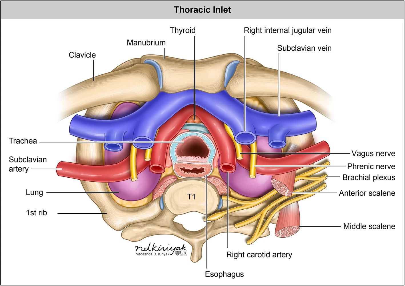
English: Anatomy of the thoracic inlet. The trachea is commonly located anterior and right lateral to the esophagus. The aortic arch typically gives rise to three main branches: the brachiocephalic artery (also known as the innominate artery or brachiocephalic trunk) which divides into the right common carotid artery and right subclavian, the left common carotid artery, and the left subclavian artery. The subclavian veins join the internal jugular veins to form the brachiocephalic veins (also known as innominate veins), which empty into the superior vena cava (SVC). The brachial plexus is comprised of nerve roots from cervical level 5 to thoracic level 1 and provides motor and sensory innervation to the shoulder and arm [1]. The phrenic nerve arises from cervical levels 3–5, running along the anterior surface of the anterior scalene muscle in the neck and enter the thorax posterior to the subclavian vein, providing innervation for the diaphragm. The bilateral vagus nerves are rarely directly discernible, even with high-resolution imaging. Their location, however, may be inferred by recognizing anatomic landmarks of their expected course and should be kept in mind during interpretation [1]. The thyroid isthmus lies just above the level of the thoracic inlet in the midline. The right and left lobes of the thyroid may extend inferiorly through the thoracic inlet into the mediastinum/substernal space [2] |
| Date | |
| Source | Nguyen, T.T., Melendez, P.E., Kaproth-Joslin, K. et al. Non-neoplastic pathology at the crossroads between neck imaging and cardiothoracic imaging. Insights Imaging 10, 116 (2019). https://doi.org/10.1186/s13244-019-0790-y |
| Author | Nadezdha D. Kiriyak |
Licensing
[edit]
This file is licensed under the Creative Commons Attribution 4.0 International license.
- You are free:
- to share – to copy, distribute and transmit the work
- to remix – to adapt the work
- Under the following conditions:
- attribution – You must give appropriate credit, provide a link to the license, and indicate if changes were made. You may do so in any reasonable manner, but not in any way that suggests the licensor endorses you or your use.
File history
Click on a date/time to view the file as it appeared at that time.
| Date/Time | Thumbnail | Dimensions | User | Comment | |
|---|---|---|---|---|---|
| current | 19:48, 9 April 2021 | | 1,418 × 1,000 (150 KB) | Balkanique (talk | contribs) | Uploaded a work by Nadezdha D. Kiriyak from Nguyen, T.T., Melendez, P.E., Kaproth-Joslin, K. et al. Non-neoplastic pathology at the crossroads between neck imaging and cardiothoracic imaging. Insights Imaging 10, 116 (2019). https://doi.org/10.1186/s13244-019-0790-y with UploadWizard |
You cannot overwrite this file.
File usage on Commons
The following page uses this file: