File:Axonal MNTB tracks in the Trapezoid Body of WT CBA mouse brainstem K Bondarenko.jpg
Original file (7,361 × 4,453 pixels, file size: 6.7 MB, MIME type: image/jpeg)
Captions
Captions
Summary[edit]
| Description |
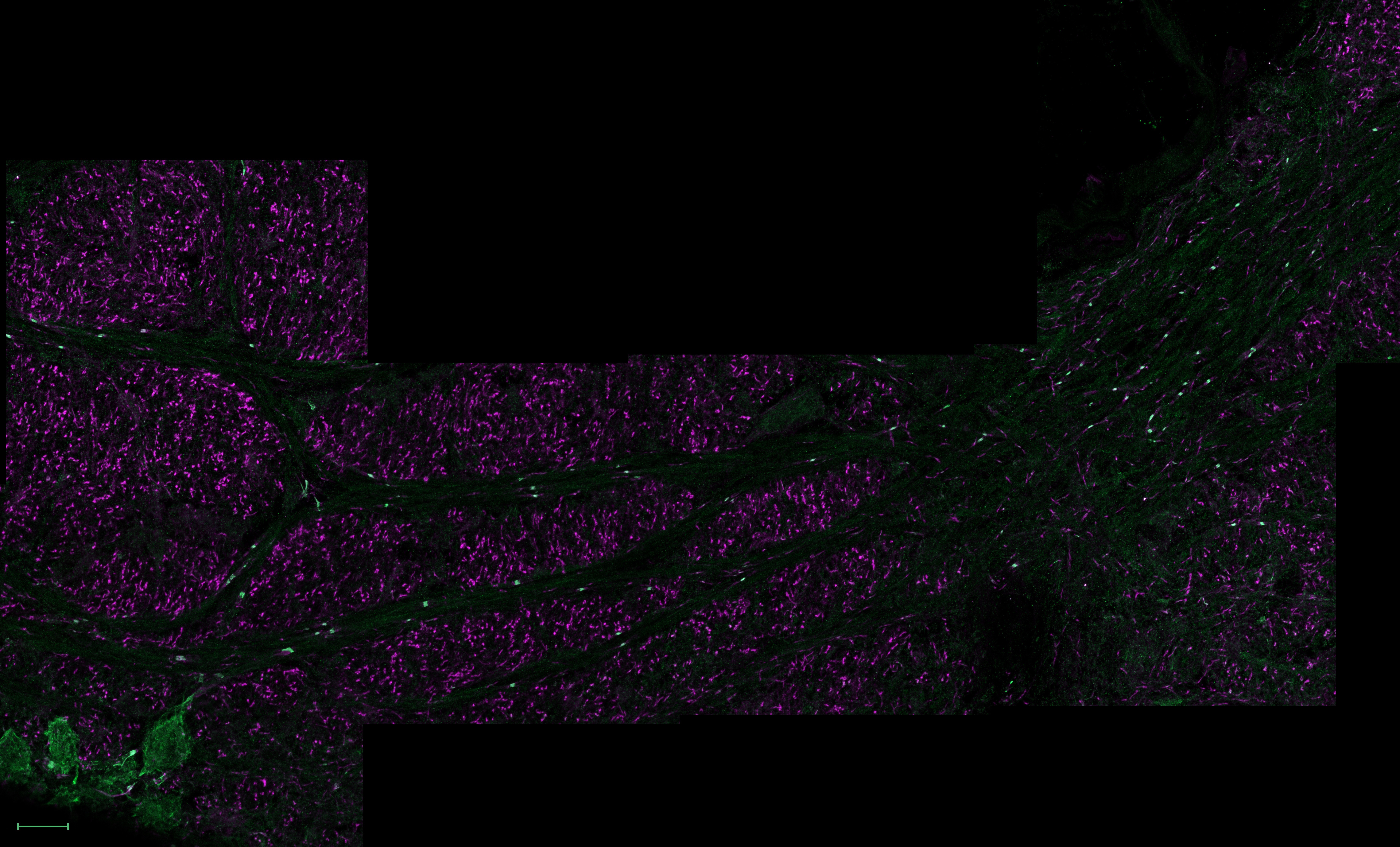
English: You look at the axonal tracks (fibres) of the Trapezoid Body in the coronal section of the mouse brainstem. The tracks originate from the bushy cells of the Cochlear Nucleus (far upper right, outside this frame) and run to the Medial Nucleus of the Trapezoid Body (MNTB, bottom left).
Voltage-gated potassium channels Kv3.1 are highlighted in green (anti-Kv3.1b + AlexaFluor488, 488 nm laser), while axon initial segments and nodes of Ranvier in magenta (anti-AnkyrinG + AlexaFluor647, 639 nm laser). The Kv3 family of voltage-gated potassium channels are important mediators of action potential repolarization. Expression of these channels enables action potentials to be of short-duration and high firing frequency. Thus, Kv3 channels play a critical role in sound processing in the brain. The big green cells at the bottom left part of the image are principal MNTB neurons. MNTB is responsible for sound localization and encoding time characteristics of complex sound, thus highly expressing Kv3.1 channels. Kv3.1 channels are also present in the nodes of Ranvier (bright green “jumpers” on the fibres of the axonal tracks with a slight magenta glow on either side), shown here by co-labelling with Ankyrin G, an intracellular adapter protein involved in the attachment of other proteins to different parts of the cell membrane. We all know that axons are long but often fail to imagine their actual span across the brain on a comprehendible scale. This image was created to highlight the fact that no information is lost while travelling encoded as an electric signal down these long wires from the ear to the centres of sound processing located deep inside the brain, and this accuracy heavily relies on Kv3.1 channels. Prep: frozen 12 µm sections from CBA WT mouse, P20. This image was acquired from several stitched single optical sections obtained using Zeiss LSM 980 confocal with a 10x objective. Scale bar: 20 µm.Українська: Ви дивитесь на аксональні доріжки (волокна) трапецієподібного тіла стовбура мозку миші (корональний зрiз). Доріжки походять від пухких клітин ядра равлика (далеко вгорі праворуч, за межами цього кадру) і йдуть до медіального ядра трапецієподібного тіла (MNTB, внизу ліворуч).
Потенціалзалежні калієві канали Kv3.1 виділені зеленим кольором (анти-Kv3.1 + AlexaFluor488, лазер 488 нм), тоді як початкові сегменти аксона та вузли Ранв’є - пурпуровим (анти-AnkyrinG + AlexaFluor647, лазер 639 нм). Сімейство потенціалзалежних калієвих каналів Kv3 є важливими медіаторами реполяризації потенціалу дії. Експресія цих каналів робить потенціали дії короткочасними з високою частотою спрацьовування. Таким чином, канали Kv3 відіграють вирішальну роль у обробці звуку в мозку. Великі зелені клітини в нижній лівій частині зображення є основними нейронами MNTB. MNTB відповідає за локалізацію звуку та часові характеристики кодування складного звуку, а отже, експресує багато каналiв Kv3.1. Канали Kv3.1 також присутні у вузлах Ранв’є (яскраво-зелені «перемички» на волокнах аксональних доріжок із легким пурпуровим світінням з обох боків), показані тут шляхом спільного позначення з Ankyrin G, що є допомагає у прикріпленні інших білків до різних частин клітинної мембрани. Ми всі знаємо, що аксони довгі, але часто не можемо уявити їх довжину у зрозумілому масштабі. Це зображення було створено для підкреслення факту, що жодної iнформацiї не втрачається, поки вона подорожує закодована як електричний сигнал по цих довгих дротах від вуха до центрів обробки звуку глибоко в мозку, i ця точнiсть досягається в тому числi завдяки Kv3.1. Препарат: заморожений зріз (12 мкм) мозку миші CBA WT (дикий тип), P19. Це зображення було отримано з кількох зшитих проекцій максимальної інтенсивності (z-стеки зображень), отриманих за допомогою конфокального мікроскопа Zeiss LSM 980 з об’єктивом 63x. Масштабна шкала: 20 мкм.English: Axonal MNTB tracks in the Trapezoid Body of WT CBA mouse brainstem |
| Date | |
| Source | Own work |
| Author | Kseniia Bondarenko |
Licensing[edit]
- You are free:
- to share – to copy, distribute and transmit the work
- to remix – to adapt the work
- Under the following conditions:
- attribution – You must give appropriate credit, provide a link to the license, and indicate if changes were made. You may do so in any reasonable manner, but not in any way that suggests the licensor endorses you or your use.
| This image was uploaded as part of Science Photo Competition 2022 in Ukraine. |
File history
Click on a date/time to view the file as it appeared at that time.
| Date/Time | Thumbnail | Dimensions | User | Comment | |
|---|---|---|---|---|---|
| current | 02:19, 19 December 2022 | | 7,361 × 4,453 (6.7 MB) | Morne Arin (talk | contribs) | Uploaded own work with UploadWizard |
You cannot overwrite this file.
File usage on Commons
The following 2 pages use this file: