File:Brain diagram without text.svg

Πρωτότυπο αρχείο (Αρχείο SVG, ονομαστικό μέγεθος 1.024 × 731 εικονοστοιχεία, μέγεθος αρχείου: 15 KB)
Λεζάντες
Λεζάντες
Σύνοψη[επεξεργασία]
| Περιγραφή |
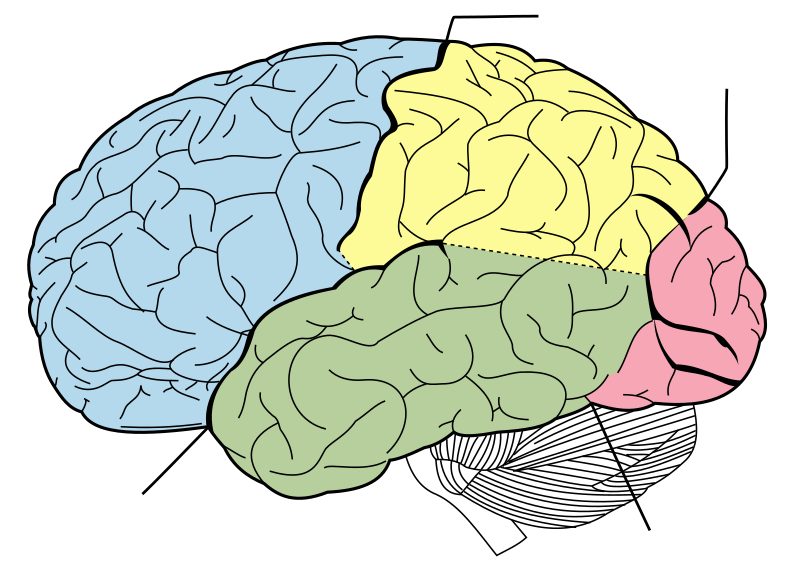
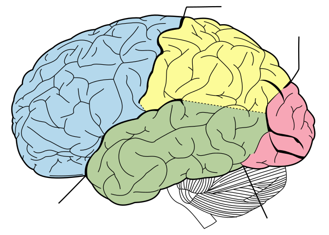
English: Principal fissures and lobes of the cerebrum viewed laterally. Principal lobes of the cerebrum viewed laterally. Figure 728 from Gray's Anatomy.
4 lines note sulci as follows
日本語: 側面から見たヒトの脳の構造(『グレイの解剖学』から引用) |
| Ημερομηνία | |
| Πηγή | Vectorized in CorelDraw by Mysid, based on the online edition of Gray's Anatomy. |
| Δημιουργός | Mysid, arrows were added by Was a bee |
| Άδεια (Επαναχρησιμοποίηση αυτού του αρχείου) |
Public domain |
| άλλες εκδόσεις |
[]
|


Αδειοδότηση[επεξεργασία]
| Unless stated otherwise, this image is from the 20th U.S. edition of Gray's Anatomy of the Human Body, originally published in 1918 and therefore lapsed into the public domain. A copy of Gray's Anatomy can be found on Bartleby and also on Yahoo!. |  |
This image is in the public domain because it is a mere mechanical scan or photocopy of a public domain original, or – from the available evidence – is so similar to such a scan or photocopy that no copyright protection can be expected to arise. The original itself is in the public domain for the following reason:
This tag is designed for use where there may be a need to assert that any enhancements (eg brightness, contrast, colour-matching, sharpening) are in themselves insufficiently creative to generate a new copyright. It can be used where it is unknown whether any enhancements have been made, as well as when the enhancements are clear but insufficient. For known raw unenhanced scans you can use an appropriate {{PD-old}} tag instead. For usage, see Commons:When to use the PD-scan tag.  | ||||
Ιστορικό αρχείου
Πατήστε σε μια ημερομηνία/ώρα για να δείτε το αρχείο όπως εμφανιζόταν εκείνη την χρονική στιγμή.
| Ημερομηνία/Ώρα | Μικρογραφία | Διαστάσεις | Χρήστης | Σχόλιο | |
|---|---|---|---|---|---|
| τρέχον | 11:36, 12 Σεπτεμβρίου 2022 | | 1.024 × 731 (15 KB) | Smasongarrison (συζήτηση | Συνεισφορά) | slimmed down with svgomg // Editing SVG source code using c:User:Rillke/SVGedit.js |
| 18:39, 10 Φεβρουαρίου 2010 |  | 1.024 × 731 (40 KB) | Was a bee (συζήτηση | Συνεισφορά) | position of arrow | |
| 09:44, 10 Φεβρουαρίου 2010 |  | 1.024 × 731 (40 KB) | Was a bee (συζήτηση | Συνεισφορά) | == {{int:filedesc}} == {{Information |Description={{en|Principal lobes of the cerebrum viewed laterally. Figure 728 from Gray's Anatomy.}}{{ja|側面から見たヒトの脳の構造(『グレイの解剖学』から引用)}} |Source=Vectorized in Cor |
Δεν μπορείτε να αντικαταστήσετε αυτό το αρχείο.
Χρήση αρχείου
Οι ακόλουθες 17 σελίδες χρησιμοποιούν προς αυτό το αρχείο:
- Lobe of the brain
- File:Brain diagram fr.png
- File:Brain diagram fr.svg
- File:Brain diagram hu.png
- File:Brain diagram it.svg
- File:Brain diagram ja.png
- File:Brain diagram ja.svg
- File:Brain diagram pl.svg
- File:Brain diagram ru.png
- File:Brain diagram without text.svg
- File:Brain diagram zh-cn.svg
- File:Gray728-ta.svg
- File:Gray728.svg
- File:Gray728 sv.svg
- File:Lobes of the brain rus.svg
- File:Lobên mejî ku.svg
- Template:Other versions/Brain diagram
Καθολική χρήση αρχείου
Τα ακόλουθα άλλα wiki χρησιμοποιούν αυτό το αρχείο:
- Χρήση σε en.wikipedia.org
- Microgyrus
- Plexus
- Neurilemma
- Suboccipital nerve
- Lesser auricular nerve
- Nucleus (neuroanatomy)
- Proisocortex
- Cingulate sulcus
- Rhinencephalon
- Paleoencephalon
- Brodmann area 37
- Brodmann area 38
- Brodmann area 39
- Sensory unit
- Hypoglossal nucleus
- Marginal nucleus of spinal cord
- Postcentral sulcus
- Supratentorial region
- Parietal-temporal-occipital
- Archicortex
- Multipolar neuron
- Trapezoid body
- Suprapineal recess
- A/S ratio
- Anterior lobe of cerebellum
- Lemniscus (anatomy)
- Cubital tunnel
- Pontine nuclei
- Laterodorsal tegmental nucleus
- Posterior grey column
- Arbor vitae (anatomy)
- Interposed nucleus
- External capsule
- Extreme capsule
- Corpora quadrigemina
- Subdural space
- Ventral root of spinal nerve
- Frontal gyri
- Galvanic vestibular stimulation
- Brodmann area 12
- Brodmann area 16
- Brodmann area 26
- Brodmann area 27
- Brodmann area 31
- Brodmann area 33
- Brodmann area 34
- Brodmann area 29
- Brodmann area 30
- Brodmann area 48
- Brodmann area 52
Δείτε περισσότερη καθολική χρήση αυτού του αρχείου.