File:Brain diagram without text.svg
跳至導覽
跳至搜尋

此 SVG 檔案的 PNG 預覽的大小:800 × 571 像素。 其他解析度:320 × 228 像素 | 640 × 457 像素 | 1,024 × 731 像素 | 1,280 × 914 像素 | 2,560 × 1,828 像素。
原始檔案 (SVG 檔案,表面大小:1,024 × 731 像素,檔案大小:15 KB)
說明
說明
添加單行說明來描述出檔案所代表的內容
摘要
[編輯]| 描述 |
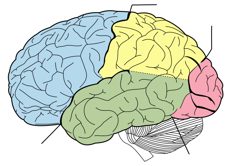
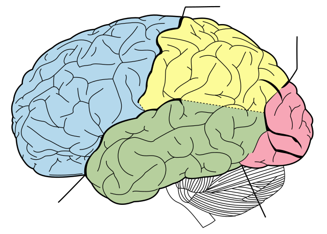
English: Principal fissures and lobes of the cerebrum viewed laterally. Principal lobes of the cerebrum viewed laterally. Figure 728 from Gray's Anatomy.
4 lines note sulci as follows
日本語: 側面から見たヒトの脳の構造(『グレイの解剖学』から引用) |
| 日期 | |
| 來源 | Vectorized in CorelDraw by Mysid, based on the online edition of Gray's Anatomy. |
| 作者 | Mysid, arrows were added by Was a bee |
| 授權許可 (重用此檔案) |
Public domain |
| 其他版本 |
[]
|


授權條款
[編輯]
| Unless stated otherwise, this image is from the 20th U.S. edition of Gray's Anatomy of the Human Body, originally published in 1918 and therefore lapsed into the public domain. A copy of Gray's Anatomy can be found on Bartleby and also on Yahoo!. |  |
本影像屬於公有領域,因為該影像是單純針對一件公有領域原作進行機械掃描,或(從現有證據來看)類似掃描而來的,或該重製照片可以預期不會有版權保護。原作屬於公有領域,是因下述原因:
本標籤主要用於該掃描件有可能使用任何增強功能(如亮度、對比度、色彩調整、銳利化等),而這些增強功能無法達到原創性而無法產生版權的情形下使用。而該標籤也能使用在無法判定本掃描件使用任何增強功能,以及已知使用增強功能但沒有充分證據時。對於採取原始忠實掃描而不使用增強功能的照片,可以適當採用{{PD-old}}標籤取代。對於本標籤的使用方法,參見如何使用PD-Scan標籤。  | ||||
檔案歷史
點選日期/時間以檢視該時間的檔案版本。
| 日期/時間 | 縮圖 | 尺寸 | 使用者 | 備註 | |
|---|---|---|---|---|---|
| 目前 | 2022年9月12日 (一) 11:36 | | 1,024 × 731(15 KB) | Smasongarrison(留言 | 貢獻) | slimmed down with svgomg // Editing SVG source code using c:User:Rillke/SVGedit.js |
| 2010年2月10日 (三) 18:39 |  | 1,024 × 731(40 KB) | Was a bee(留言 | 貢獻) | position of arrow | |
| 2010年2月10日 (三) 09:44 |  | 1,024 × 731(40 KB) | Was a bee(留言 | 貢獻) | == {{int:filedesc}} == {{Information |Description={{en|Principal lobes of the cerebrum viewed laterally. Figure 728 from Gray's Anatomy.}}{{ja|側面から見たヒトの脳の構造(『グレイの解剖学』から引用)}} |Source=Vectorized in Cor |
無法覆蓋此檔案。
檔案用途
下列17個頁面有用到此檔案:
- Lobe of the brain
- File:Brain diagram fr.png
- File:Brain diagram fr.svg
- File:Brain diagram hu.png
- File:Brain diagram it.svg
- File:Brain diagram ja.png
- File:Brain diagram ja.svg
- File:Brain diagram pl.svg
- File:Brain diagram ru.png
- File:Brain diagram without text.svg
- File:Brain diagram zh-cn.svg
- File:Gray728-ta.svg
- File:Gray728.svg
- File:Gray728 sv.svg
- File:Lobes of the brain rus.svg
- File:Lobên mejî ku.svg
- Template:Other versions/Brain diagram
全域檔案使用狀況
以下其他 wiki 使用了這個檔案:
- en.wikipedia.org 的使用狀況
- Microgyrus
- Plexus
- Neurilemma
- Suboccipital nerve
- Lesser auricular nerve
- Nucleus (neuroanatomy)
- Proisocortex
- Cingulate sulcus
- Rhinencephalon
- Paleoencephalon
- Brodmann area 37
- Brodmann area 38
- Brodmann area 39
- Sensory unit
- Hypoglossal nucleus
- Marginal nucleus of spinal cord
- Postcentral sulcus
- Supratentorial region
- Parietal-temporal-occipital
- Archicortex
- Multipolar neuron
- Trapezoid body
- Suprapineal recess
- A/S ratio
- Anterior lobe of cerebellum
- Lemniscus (anatomy)
- Cubital tunnel
- Pontine nuclei
- Laterodorsal tegmental nucleus
- Posterior grey column
- Arbor vitae (anatomy)
- Interposed nucleus
- External capsule
- Extreme capsule
- Corpora quadrigemina
- Subdural space
- Ventral root of spinal nerve
- Frontal gyri
- Galvanic vestibular stimulation
- Brodmann area 12
- Brodmann area 16
- Brodmann area 26
- Brodmann area 27
- Brodmann area 31
- Brodmann area 33
- Brodmann area 34
- Brodmann area 29
- Brodmann area 30
- Brodmann area 48
- Brodmann area 52
檢視此檔案的更多全域使用狀況。