File:Cel Beta Electofisiol Unica.png
Jump to navigation
Jump to search
Size of this preview: 800 × 330 pixels. Other resolutions: 320 × 132 pixels | 640 × 264 pixels | 1,024 × 422 pixels | 2,028 × 836 pixels.
Original file (2,028 × 836 pixels, file size: 305 KB, MIME type: image/png)
Captions
Captions
Add a one-line explanation of what this file represents
Summary
[edit]
| Description |
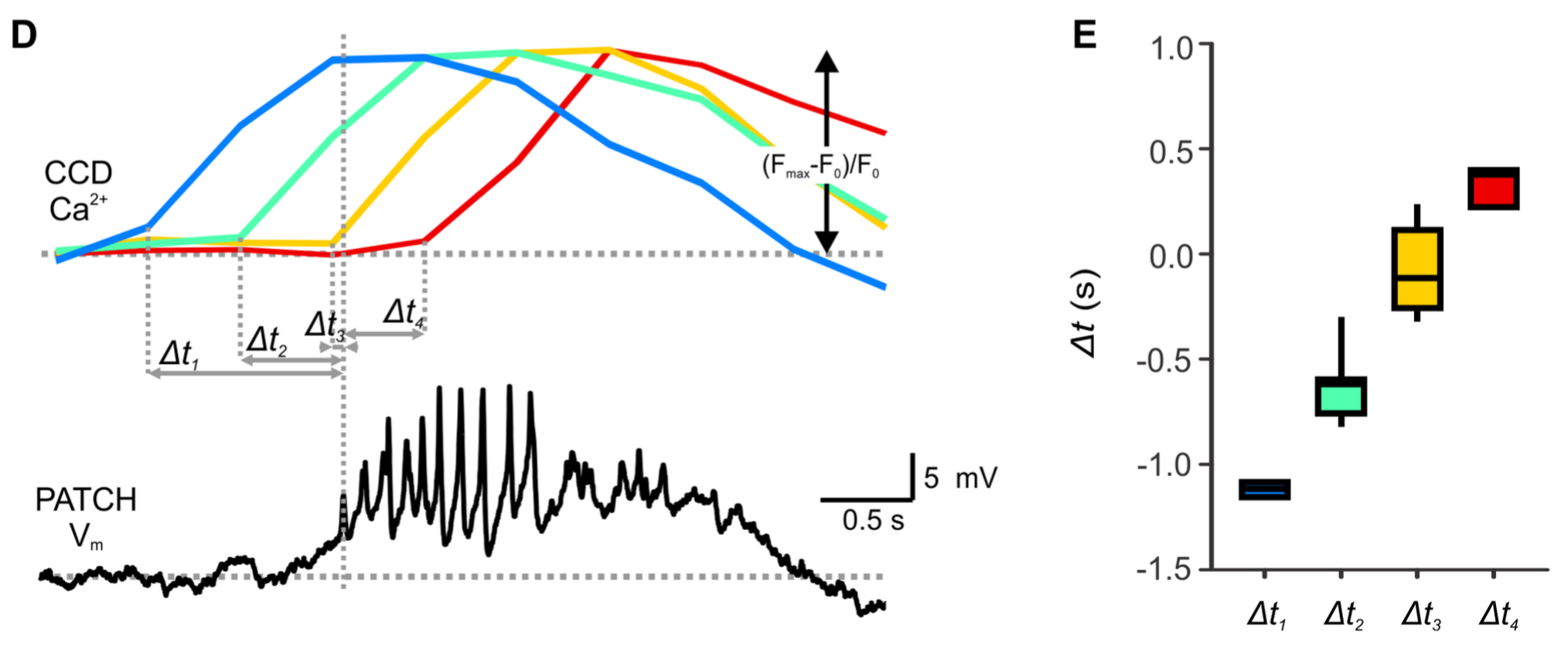
Español: Figura 1. Registro simultáneo de las oscilaciones de potencial de membrana que se producen en la célula sujetada con parche y las oscilaciones de calcio intracelular [Ca 2+ ] i que se producen en otras células del mismo islote de Langerhans. D. Una representación más detallada del área indicada en C. El inicio de los picos de alta frecuencia en la célula parcheada se correlaciona temporalmente mejor con el inicio de la oscilación [Ca 2+] i en el grupo de células indicado en naranja. Para este grupo de células, la distancia al origen de la oscilación era aproximadamente la misma que para la célula parcheada. |
| Date | |
| Source | The Relationship between Membrane Potential and Calcium Dynamics in Glucose-Stimulated Beta Cell Syncytium in Acute Mouse Pancreas Tissue Slices. (2013). PLoS ONE 8(12): e82374. doi:10.1371/journal.pone.0082374 |
| Author | Jurij Dolenšek, Andraž Stožer, Maša Skelin Klemen, Evan W. Miller, Marjan Slak Rupnik |
Licensing
[edit]
This file is licensed under the Creative Commons Attribution 4.0 International license.
- You are free:
- to share – to copy, distribute and transmit the work
- to remix – to adapt the work
- Under the following conditions:
- attribution – You must give appropriate credit, provide a link to the license, and indicate if changes were made. You may do so in any reasonable manner, but not in any way that suggests the licensor endorses you or your use.
File history
Click on a date/time to view the file as it appeared at that time.
| Date/Time | Thumbnail | Dimensions | User | Comment | |
|---|---|---|---|---|---|
| current | 16:46, 18 April 2023 | 2,028 × 836 (305 KB) | Sanador2.0 (talk | contribs) | Uploaded a work by Jurij Dolenšek, Andraž Stožer, Maša Skelin Klemen, Evan W. Miller, Marjan Slak Rupnik from The Relationship between Membrane Potential and Calcium Dynamics in Glucose-Stimulated Beta Cell Syncytium in Acute Mouse Pancreas Tissue Slices. (2013). PLoS ONE 8(12): e82374. doi:10.1371/journal.pone.0082374 with UploadWizard |
You cannot overwrite this file.
File usage on Commons
There are no pages that use this file.
File usage on other wikis
The following other wikis use this file:
- Usage on es.wikipedia.org