File:Chorioamnionitis - intermed mag.jpg
跳至導覽
跳至搜尋

預覽大小:800 × 533 像素。 其他解析度:320 × 213 像素 | 640 × 427 像素 | 1,024 × 683 像素 | 1,280 × 853 像素 | 2,560 × 1,707 像素 | 4,272 × 2,848 像素。
原始檔案 (4,272 × 2,848 像素,檔案大小:4.62 MB,MIME 類型:image/jpeg)
說明
說明
添加單行說明來描述出檔案所代表的內容
摘要
[編輯]| 描述 |
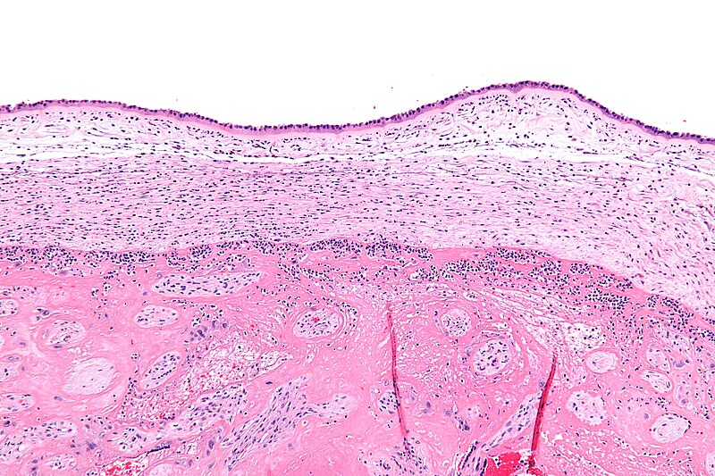
English: Intermediate magnification micrograph chorioamnionitis. H&E stain.
The amnion is seen at the very top of the image and composed of a simple cuboidal epithelium and a layer of eosinophilic (pink) connective tissue. It has a few scattered neutrophils - which makes the diagnosis of chorioamnionitis. Below the amnion is a cleft and then the chorion, which also has neutrophils. The fetus (baby) - not shown - would be below the image (and surrounded by amnionic fluid). The pattern of inflammation seen in this image is typical of chorioamnionitis; most chorioamnionitis results from an ascending infection, i.e. the microorganisms ascend from the vagina/cervix. Related images
|
| 來源 | 自己的作品 |
| 作者 | Nephron |


授權條款
[編輯]
我,本作品的著作權持有者,決定用以下授權條款發佈本作品:
此檔案採用創用CC 姓名標示-相同方式分享 3.0 未在地化版本授權條款。
- 您可以自由:
- 分享 – 複製、發佈和傳播本作品
- 重新修改 – 創作演繹作品
- 惟需遵照下列條件:
- 姓名標示 – 您必須指名出正確的製作者,和提供授權條款的連結,以及表示是否有對內容上做出變更。您可以用任何合理的方式來行動,但不得以任何方式表明授權條款是對您許可或是由您所使用。
- 相同方式分享 – 如果您利用本素材進行再混合、轉換或創作,您必須基於如同原先的相同或兼容的條款,來分布您的貢獻成品。

|
已授權您依據自由軟體基金會發行的無固定段落、封面文字和封底文字GNU自由文件授權條款1.2版或任意後續版本,對本檔進行複製、傳播和/或修改。該協議的副本列在GNU自由文件授權條款中。 |
您可以選擇您需要的授權條款。
檔案歷史
點選日期/時間以檢視該時間的檔案版本。
| 日期/時間 | 縮圖 | 尺寸 | 使用者 | 備註 | |
|---|---|---|---|---|---|
| 目前 | 2011年8月20日 (六) 04:06 | | 4,272 × 2,848(4.62 MB) | Nephron(留言 | 貢獻) | {{Information |Description ={{en|1=Intermediate magnification micrograph '''chorioamnionitis'''. H&E stain. The amnion is seen at the very top of the image and composed of a [[w:simp |
無法覆蓋此檔案。
檔案用途
下列5個頁面有用到此檔案:
全域檔案使用狀況
以下其他 wiki 使用了這個檔案:
- de.wikibooks.org 的使用狀況
- en.wikipedia.org 的使用狀況
- es.wikipedia.org 的使用狀況
- fa.wikipedia.org 的使用狀況
- hy.wikipedia.org 的使用狀況
- tr.wikipedia.org 的使用狀況
- zh.wikipedia.org 的使用狀況