File:Crystal Structure of the FokI endonuklease - PDB-2FOK.png
Jump to navigation
Jump to search
Size of this preview: 768 × 600 pixels. Other resolutions: 307 × 240 pixels | 615 × 480 pixels | 983 × 768 pixels | 1,280 × 1,000 pixels.
Original file (1,280 × 1,000 pixels, file size: 696 KB, MIME type: image/png)
Captions
Captions
Add a one-line explanation of what this file represents
Summary
[edit]
| Description |
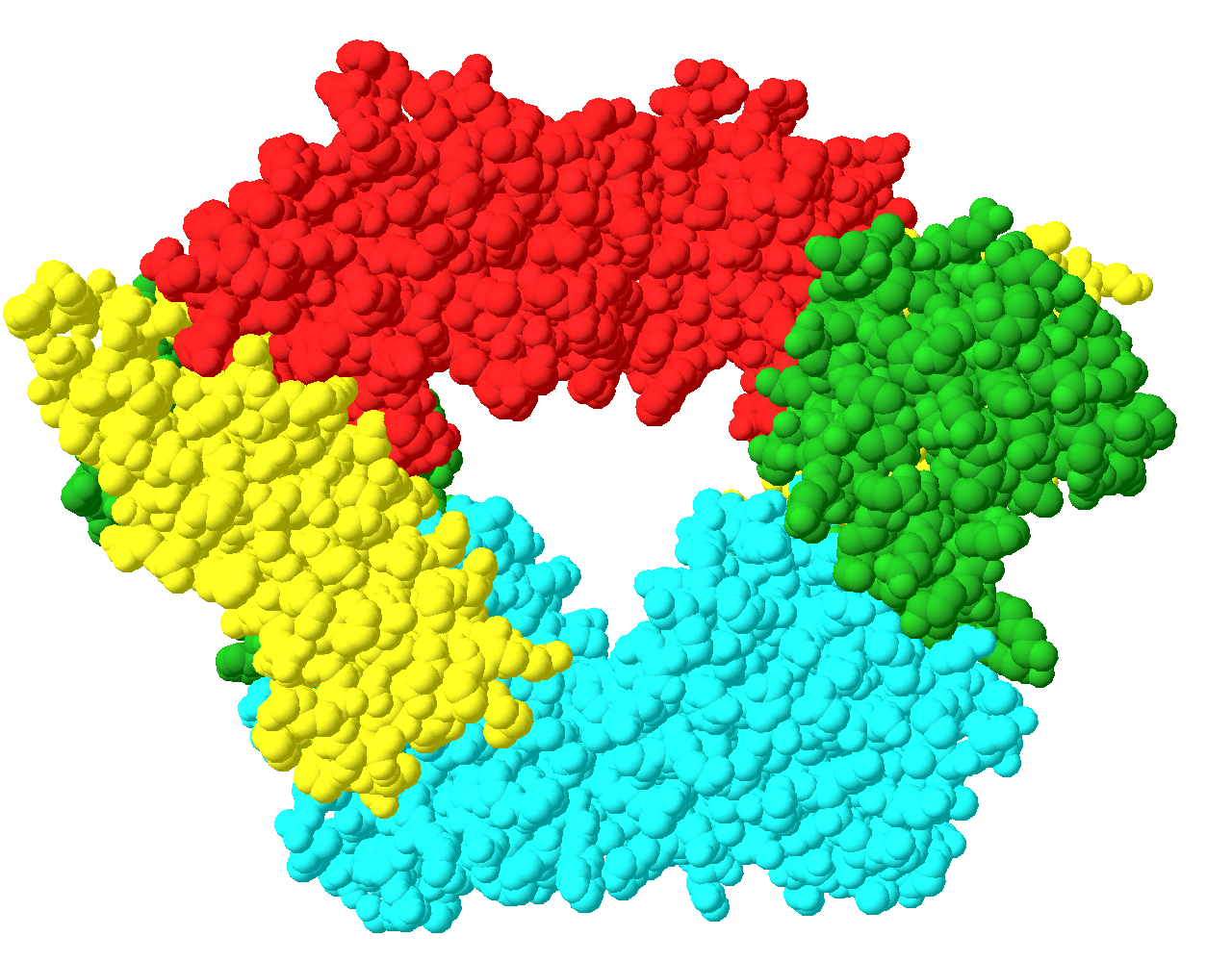
English: Endonuclease FokI dimer derived from Planomicrobium okeanokoites. Color was determined individual domain name:
- red (the first domain; D1), green (second domain; D2), yellow (third domain; D3) - responsible for the connection to DNA Endonuclease Polski: Dimer endonukleazy FokI pochodzącej z Planomicrobium okeanokoites. Kolorami oznaczono poszczególne domeny:
- czerwony (pierwsza domena; D1), zielony (druga domena; D2), żółty (trzecia domena; D3) - odpowiadające za przyłączenie endonukleazy do nici DNA |
| Date | |
| Source | Own work |
| Author | Marcello002 |
Licensing
[edit]
| I, the copyright holder of this work, release this work into the public domain. This applies worldwide. In some countries this may not be legally possible; if so: I grant anyone the right to use this work for any purpose, without any conditions, unless such conditions are required by law. |
File history
Click on a date/time to view the file as it appeared at that time.
| Date/Time | Thumbnail | Dimensions | User | Comment | |
|---|---|---|---|---|---|
| current | 13:12, 27 December 2009 | | 1,280 × 1,000 (696 KB) | Marcello002 (talk | contribs) | {{Information |Description={{en|1=Endonuclease FokI dimer derived from ''Planomicrobium okeanokoites''. Color was determined individual domain name: - red (the first domain; D1), green (second domain; D2), yellow (third domain; D3) - responsible for the c |
You cannot overwrite this file.
File usage on Commons
The following page uses this file:
- File:Crystal Structure of the FokI endonuklease - PDB-2FOK .png (file redirect)
File usage on other wikis
The following other wikis use this file:
- Usage on pl.wikipedia.org