File:EB1911 Flagellata (2).jpg

Original file (776 × 1,701 pixels, file size: 563 KB, MIME type: image/jpeg)
Captions
Captions
Summary
[edit]| Description |
English: Flagellata:
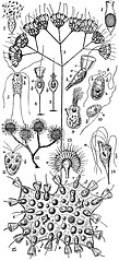
1. Salpingoeca fusiformis, S. Kent (Choanoflagellata). The protoplasmic body is drawn together within the goblet-shaped shell, and divided into numerous spores. 2. Escape of the spores of the same as monoflagellate and swarm-spores. 3. Codosiga umbellata, Tatem (Choanoflagellata); adult colony formed by dichotomous growth. 4. A single zooid of the same. a = nucleus. b = contractile vacuole. c = the characteristic “collar” of naked streaming protoplasm. 5. Hexamita inflata, Duj.(Distomatidae); normal adult. 6, 7 Salpingoeca urceolata, S Kent (Choanoflagellata)—6, with collar extended; 7, with collar retracted within the stalked cup. 8 Polytoma uvella, Mull. sp. (Chlamydomonadidae). 9. Lophomonas blattarum, Stein (Trichonymphidae) from the intestine of Blatta orientalis. 10. Bodolens, Mull. (Bodonidae), the wavy filament is a tractellum, the straight one is a trailing thread. 11. Tetramitus sulcatus, Stein (Tetramitidae) 12. Anthophysa vegetans, O.F. Müller (Monadidae). A typical, erect, shortly-branching colony stock with four terminal monad-clusters. 13. Monad cluster of the same in optical section, showing the relation of the individual monads or flagellate zooids to the stem d. 14. Tetramitus rostratus, Perty (Tetramitidae). a = nucleus. b = contractile vacuole. 15. Proterospongia Haeckeli, Saville Kent (Choanoflagellata); A social colony of about forty flagellate zooids. a = nucleus. b = contractile vacuole. c = amoebiform cell sunk within the colonial gelatinous test compared by S. Kent to a mesoderm cell of the sponges. d = similar cell reproducing by transverse fission. e = normal cells, with their collars contracted. f = substance of test.g = individual reproducing by multiple fission, producing microzoospores, comparable to the spermatozoa of sponges. |
|||
| Date | published 1911 | |||
| Source | “Flagellata,” Encyclopædia Britannica (11th ed.), v. 10, 1911, p. 466, fig. 2. | |||
| Author | Unknown artist | |||
| Permission (Reusing this file) |
|

.jpg&action=edit§ion=1)
File history
Click on a date/time to view the file as it appeared at that time.
| Date/Time | Thumbnail | Dimensions | User | Comment | |
|---|---|---|---|---|---|
| current | 16:05, 8 August 2018 | | 776 × 1,701 (563 KB) | Bob Burkhardt (talk | contribs) | {{Information |description ={{en|1=Flagellata: 1. Salpingoeca fusiformis, S. Kent (Choanoflagellata). The protoplasmic body is drawn together within the goblet-shaped shell, and divided into numerous spores. 2. Escape of the spores of the same as monoflagellate and swarm-spores. 3. Codosiga umbellata, Tatem (Choanoflagellata); adult colony formed by dichotomous growth. 4. A single zooid of the same. a = nucleus. b = contractile vacuole. c = the characteristic “collar” of na... |
You cannot overwrite this file.
File usage on Commons
There are no pages that use this file.
File usage on other wikis
The following other wikis use this file:
- Usage on en.wikisource.org
.jpg&oldid=743524110)