File:EB1911 Lamellibranchia - ctenidia of Nucula.jpg
Jump to navigation
Jump to search
Size of this preview: 401 × 599 pixels. Other resolutions: 160 × 240 pixels | 321 × 480 pixels | 747 × 1,116 pixels.
Original file (747 × 1,116 pixels, file size: 311 KB, MIME type: image/jpeg)
Captions
Captions
Add a one-line explanation of what this file represents
Summary
[edit]| Description |
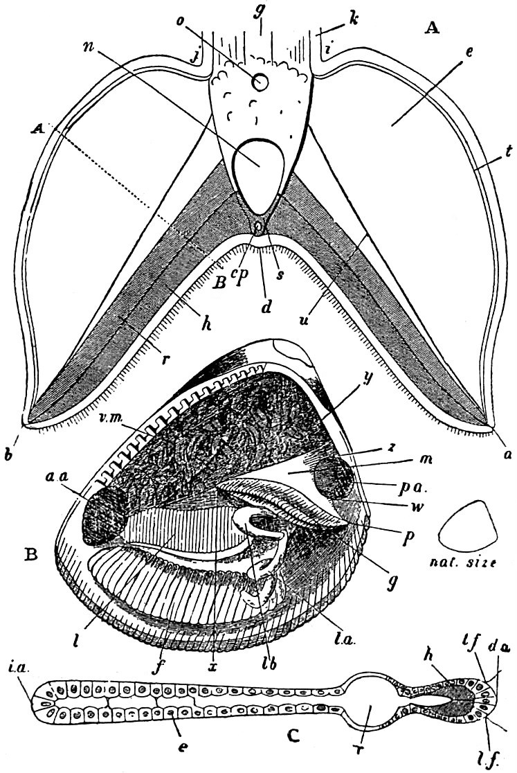
English: Structure of the ctenidia of Nucula. See legend below. |
|||
| Date | published 1911 | |||
| Source | “Lamellibranchia,” Encyclopædia Britannica (11th ed.), v. 16, 1911, p. 116, fig. 10. | |||
| Author | After Mitsukuri. | |||
| Permission (Reusing this file) |
|

English: Legend:
| A. | Section across the axis of a ctenidium with a pair of plates—flattened and shortened filaments—attached. |
| i, j, k, g, | Are placed on or near the membrane which attaches the axis of the ctenidium to the side of the body. |
| a, b, | Free extremities of the plates (filaments). |
| d, | Mid-line of the inferior border. |
| e, | Surface of the plate. |
| t, | Its upper border. |
| h, | Chitinous lining of the plate. |
| r, | Dilated blood-space. |
| u, | Fibrous tract. |
| o, | Upper blood-vessel of the axis. |
| n, | Lower blood-vessel of the axis. |
| s, | Chitinous framework of the axis. |
| cp, | Canal in the same. |
| A, B, | Line along which the cross-section C of the plate is taken. |
| B. | Animal of a male Nucula proxima, Say, as seen when the left valve of the shell and the left half of the mantle-skirt are removed. |
| a, a, | Anterior adductor muscle. |
| p.a, | Posterior adductor muscle. |
| v.m, | Visceral mass. |
| f, | Foot. |
| g, | Gill. |
| l, | Labial Tentacle. |
| l.a, | Filamentous appendage of the labial tentacle. |
| lb, | Hood-like appendage of the labial tentacle. |
| m, | Membrane suspending the gill and attached to the body along the line x, y, z, w. |
| p, | Posterior end of the gill (ctenidium). |
| C. | Section across one of the gill-plates (A, B, in A) comparable with fig. 11 C. |
| i.a, | Outer border. |
| d.a, | Axial border. |
| l.f, | Latero-frontal epithelium. |
| e, | Epithelium of general surface. |
| r, | Dilated blood-space. |
| h, | Chitinous lining (compare A). |
File history
Click on a date/time to view the file as it appeared at that time.
| Date/Time | Thumbnail | Dimensions | User | Comment | |
|---|---|---|---|---|---|
| current | 16:26, 8 April 2019 | | 747 × 1,116 (311 KB) | Bob Burkhardt (talk | contribs) | {{Information |description ={{en|1=Structure of the ctenidia of ''Nucula''.}} |date =published 1911 |source =“Lamellibranchia,” ''Encyclopædia Britannica'' (11th ed.), v. 16, 1911, p. 116, fig. 10. |author =After Mitsukuri. |permission ={{PD-Britannica}} }} Category:Nucula Category:Mollusca anatomy |
You cannot overwrite this file.
File usage on Commons
The following page uses this file:
File usage on other wikis
The following other wikis use this file:
- Usage on en.wikisource.org