File:Embryonic Development CNS ar.png
From Wikimedia Commons, the free media repository
Jump to navigation
Jump to search
Embryonic_Development_CNS_ar.png (350 × 325 pixels, file size: 446 KB, MIME type: image/png)
File information
Structured data
Captions
Captions
Embryonic Development central nervous system
التطور الجنيني للجهاز العصبي المركزي
Summary[edit]
| DescriptionEmbryonic Development CNS ar.png |
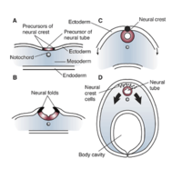
العربية: تكون الأنبوب العصبي (رؤية عرضية). في مرحلة مبكرة لنمو الجنين، يقوم شريط من الخلايا المتخصصة يسمى الحبل الظهري (A) بتحفيز خلايا الأديم الظاهر مباشرةً فوقه لتصبح الجهاز العصبي الأولي (أي الظهارة العصبية). ثم تتخدَّد الظهارة العصبية وتثنى فوق (B). عندما تندمج أطراف الطيات معًا، يتشكل أنبوب مجوف (أي الأنبوب العصبي) (C) -مقدمة الدماغ والحبل الشوكي. وفي الوقت نفسه، يستمر الأديم الظاهر والأديم الباطني في الانحناء والاندماج تحت الجنين لإنشاء تجويف الجسم، لإكمال تحول الجنين من قرص مسطح إلى جسم ثلاثي الأبعاد. تهاجر الخلايا الناشئة من الأطراف الملتحمة للأديم الظاهر العصبي (أي خلايا العرف العصبي) إلى مواقع مختلفة في جميع أنحاء الجنين، حيث تبدأ في تطوير هياكل الجسم المتنوعة (D). قام الباحثون الذين يدرسون متلازمة الجنين الكحولي بدراسة خلايا العرف العصبي على نطاق واسع، لأنها حساسة بشكل خاص للإصابة الناجمة عن الكحول وموت الخلايا.
English: Formation of the neural tube (cross view). Early in an embryo’s development, a strip of specialized cells called the notochord (A) induces the cells of the ectoderm directly above it to become the primitive nervous system (i.e., neuroepithelium). The neuroepithelium then wrinkles and folds over (B). As the tips of the folds fuse together, a hollow tube (i.e., the neural tube) forms (C)—the precursor of the brain and spinal cord. Meanwhile, the ectoderm and endoderm continue to curve around and fuse beneath the embryo to create the body cavity, completing the transformation of the embryo from a flattened disk to a three–dimensional body. Cells originating from the fused tips of the neuroectoderm (i.e., neural crest cells) migrate to various locations throughout the embryo, where they will initiate the development of diverse body structures (D). Researchers investigating fetal alcohol syndrome have extensively studied neural crest cells, because they are particularly sensitive to alcohol–induced injury and cell death. |
| Date | |
| Source | Derivative from this file |
| Author | |
| Other versions |
This file was derived from: Embryonic Development CNS.png:  |
| This is a retouched picture, which means that it has been digitally altered from its original version. Modifications: Translated to Arabic - عُرِبَت. The original can be viewed here: Embryonic Development CNS.png:
|
Licensing[edit]
I, the copyright holder of this work, hereby publish it under the following license:
This file is licensed under the Creative Commons Attribution-Share Alike 4.0 International license.
- You are free:
- to share – to copy, distribute and transmit the work
- to remix – to adapt the work
- Under the following conditions:
- attribution – You must give appropriate credit, provide a link to the license, and indicate if changes were made. You may do so in any reasonable manner, but not in any way that suggests the licensor endorses you or your use.
- share alike – If you remix, transform, or build upon the material, you must distribute your contributions under the same or compatible license as the original.
File history
Click on a date/time to view the file as it appeared at that time.
| Date/Time | Thumbnail | Dimensions | User | Comment | |
|---|---|---|---|---|---|
| current | 07:51, 28 May 2021 |  | 350 × 325 (446 KB) | لوقا (talk | contribs) | Uploaded own work with UploadWizard |
You cannot overwrite this file.
File usage on Commons
The following page uses this file:
File usage on other wikis
The following other wikis use this file:
- Usage on ar.wikipedia.org
Metadata
This file contains additional information such as Exif metadata which may have been added by the digital camera, scanner, or software program used to create or digitize it. If the file has been modified from its original state, some details such as the timestamp may not fully reflect those of the original file. The timestamp is only as accurate as the clock in the camera, and it may be completely wrong.
| Horizontal resolution | 28.35 dpc |
|---|---|
| Vertical resolution | 28.35 dpc |
| File change date and time | 07:46, 28 May 2021 |
