File:Fimmu-09-03042-g001 (1).jpg
Jump to navigation
Jump to search

Size of this preview: 800 × 566 pixels. Other resolutions: 320 × 226 pixels | 640 × 453 pixels | 1,084 × 767 pixels.
Original file (1,084 × 767 pixels, file size: 824 KB, MIME type: image/jpeg)
Captions
Captions
Schistosomiasis
Summary[edit]
.jpg&action=edit§ion=1)
| Description |
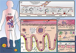
English: Figure 1. An overview of S. mansoni egg migration. Schistosome egg transit is facilitated by a series of host interactions at the intestinal and vascular interface. (A) The development of schistosomes into sexually mature, egg-producing adults occurs within the portal vein (~3–5 weeks post infection) and requires the transduction of host-derived signals (including those from the innate and adaptive immune system) to the developing worm pair. Once sexual maturity is reached, worm pairs migrate toward the mesenteric vessels, where the females lay approximately 300 eggs per day and actively modulates the intravascular environment to support their long-term survival. The production of eggs at ~5–6 weeks post infection is a milestone event in the schistosome life cycle, that is characterized by induction of a marked Th2 response and angiogenesis. Notably, the generation of a Th2 response by the host is critical for egg passage, and new vessel formation may favor egg transit, promoting the recruitment of immune cells and nutrients to developing granulomas. Freshly deposited eggs cannot move by themselves and must somehow attach and extravasate the endothelium. Although yet to be fully defined, this process may involve E-selectin:-Lewis-x interactions, and participation from platelets, ICAM-1 and VCAM-1. While a large proportion of eggs successfully penetrate the endothelium and reach intestinal tissue, many are swept to the liver or other distal locations (e.g., brain or spinal cord). Since schistosome eggs are unable to transit through these organs, overwhelming tissue pathology and inflammation may ensue. (B) Once schistosome eggs have passed across the host endothelium and out of the vasculature, they must cross the multi-layered intestinal wall. The host immune system responds to transiting eggs via an inflammatory granuloma response, in which individual eggs are encapsulated by immune cells [including alternatively activated (AA) macrophages, Th2 cells and eosinophils] and extracellular matrix (ECM), which protects host tissues from egg-derived toxins, but ultimately leads to formation of fibrotic lesions. For unknown reasons, granulomatous responses need to successfully develop for effective egg excretion from the host. Accordingly, schistosomes and their host have co-evolved a wide range of mechanisms to skew the host immune response toward granuloma-inducing Th2 profile. These include the ability of soluble egg antigens (SEA) to promote alternative activation in macrophages and to condition dendritic cells (DCs) for Th2 polarization. However, to prevent unwanted bystander tissue damage and potentially fatal immunopathology, schistosomes also implement various strategies to dampen host immunity and expanded regulatory networks (Bregs and Tregs). There remain many unknowns surrounding egg migration. This includes the molecules secreted by eggs to disrupt host barriers and modulate immune responses and, importantly, how egg penetration and intestinal ‘leakiness' may influence local and systemic immune reactions. |
| Date | |
| Source | https://www.frontiersin.org/journals/immunology/articles/10.3389/fimmu.2018.03042/full |
| Author | Alice H. Costain,Andrew S. MacDonald,Hermelijn H. Smits |
Licensing[edit]
.jpg&action=edit§ion=2)
This file is licensed under the Creative Commons Attribution 4.0 International license.
- You are free:
- to share – to copy, distribute and transmit the work
- to remix – to adapt the work
- Under the following conditions:
- attribution – You must give appropriate credit, provide a link to the license, and indicate if changes were made. You may do so in any reasonable manner, but not in any way that suggests the licensor endorses you or your use.
File history
Click on a date/time to view the file as it appeared at that time.
| Date/Time | Thumbnail | Dimensions | User | Comment | |
|---|---|---|---|---|---|
| current | 23:43, 21 April 2024 | | 1,084 × 767 (824 KB) | Ozzie10aaaa (talk | contribs) | Uploaded a work by Alice H. Costain,Andrew S. MacDonald,Hermelijn H. Smits from https://www.frontiersin.org/journals/immunology/articles/10.3389/fimmu.2018.03042/full with UploadWizard |
You cannot overwrite this file.
File usage on Commons
There are no pages that use this file.
.jpg&oldid=876273545)