File:Fluorescence.microscope2.jpg
From Wikimedia Commons, the free media repository
Jump to navigation
Jump to search

Size of this preview: 588 × 599 pixels. Other resolutions: 235 × 240 pixels | 471 × 480 pixels | 753 × 768 pixels | 1,005 × 1,024 pixels | 2,428 × 2,475 pixels.
Original file (2,428 × 2,475 pixels, file size: 1.21 MB, MIME type: image/jpeg)
File information
Structured data
Captions
Captions
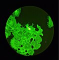
COS-7 cells colored with phalloidin under fluorescence microscope.
Cellule COS-7 colorate con falloidina e osservate con microscopio a fluorescenza.
Summary
[edit]| DescriptionFluorescence.microscope2.jpg |
English: A cell culture (COS-7) colored with phalloidin-fitc (phalloidin conjugated to fluorescein diluted in PBS), a compound which binds to actine proteins within the cytoskeleton, and colours them a bright green when observed using a fluorescence microscope.
Italiano: Cultura cellulare (COS-7) colorata con falloidina-fitc (falloidina coniugata a fluoresceina diluita in PBS), un composto che si lega all'actina presente nel citoscheletro e la colora di un verde brillante quando osservata usando un microscopio a fluorescenza. |
| Date | |
| Source | Own work |
| Author | Bioguy10 |
Licensing
[edit]I, the copyright holder of this work, hereby publish it under the following license:
| This file is made available under the Creative Commons CC0 1.0 Universal Public Domain Dedication. | |
| The person who associated a work with this deed has dedicated the work to the public domain by waiving all of their rights to the work worldwide under copyright law, including all related and neighboring rights, to the extent allowed by law. You can copy, modify, distribute and perform the work, even for commercial purposes, all without asking permission.
http://creativecommons.org/publicdomain/zero/1.0/deed.enCC0Creative Commons Zero, Public Domain Dedicationfalsefalse |
| This file was uploaded as part of Wiki Science Competition 2023. |
File history
Click on a date/time to view the file as it appeared at that time.
| Date/Time | Thumbnail | Dimensions | User | Comment | |
|---|---|---|---|---|---|
| current | 01:31, 12 December 2023 |  | 2,428 × 2,475 (1.21 MB) | Bioguy10 (talk | contribs) | Uploaded own work with UploadWizard |
You cannot overwrite this file.
File usage on Commons
The following 5 pages use this file:
Metadata
This file contains additional information such as Exif metadata which may have been added by the digital camera, scanner, or software program used to create or digitize it. If the file has been modified from its original state, some details such as the timestamp may not fully reflect those of the original file. The timestamp is only as accurate as the clock in the camera, and it may be completely wrong.
| Camera manufacturer | samsung |
|---|---|
| Camera model | SM-S906B |
| Date and time of data generation | 16:46, 9 November 2022 |
| Exposure time | 1/10 sec (0.1) |
| F-number | f/1.8 |
| ISO speed rating | 1,250 |
| User comments | |
| Lens focal length | 5.4 mm |
| Compression scheme | JPEG (old) |
| Offset to JPEG SOI | 956 |
| Bytes of JPEG data | 10,521 |
| Height | 3,000 px |
| Orientation | Normal |
| File change date and time | 16:46, 9 November 2022 |
| Vertical resolution | 72 dpi |
| Horizontal resolution | 72 dpi |
| Width | 4,000 px |
| Software used | S906BXXU2BVJA |
| Y and C positioning | Centered |
| APEX aperture | 1.69 |
| Exif version | 2.2 |
| Scene type | 63 |
| APEX exposure bias | 0 |
| Exposure Program | Normal program |
| Color space | sRGB |
| Unique image ID | D50XLOD00PM |
| Maximum land aperture | 1.69 APEX (f/1.8) |
| APEX brightness | −3.69 |
| Supported Flashpix version | 1 |
| DateTimeOriginal subseconds | 0546 |
| White balance | Auto white balance |
| Custom image processing | Normal process |
| Exposure mode | Auto exposure |
| Flash | Flash did not fire |
| DateTime subseconds | 0546 |
| Saturation | Normal |
| Meaning of each component |
|
| Focal length in 35 mm film | 23 mm |
| Contrast | Normal |
| Sharpness | Normal |
| DateTimeDigitized subseconds | 0546 |
| Digital zoom ratio | 0 |
| Date and time of digitizing | 16:46, 9 November 2022 |
| APEX shutter speed | 3.32 |
| Metering mode | Center weighted average |
| Scene capture type | Standard |
| Light source | Unknown |
