File:Fmicb-13-818658-g001.jpg
Jump to navigation
Jump to search

Size of this preview: 800 × 479 pixels. Other resolutions: 320 × 191 pixels | 640 × 383 pixels | 1,024 × 613 pixels | 1,280 × 766 pixels | 2,560 × 1,532 pixels | 4,317 × 2,583 pixels.
Original file (4,317 × 2,583 pixels, file size: 540 KB, MIME type: image/jpeg)
Captions
Captions
ocular herpes
Summary
[edit]
| Description |
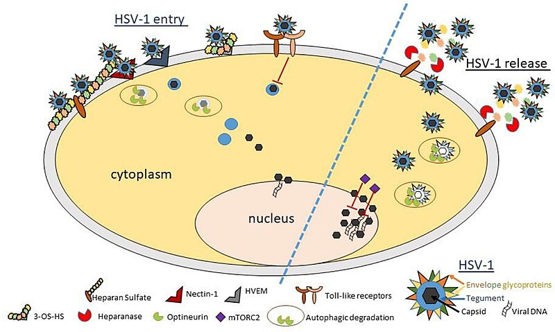
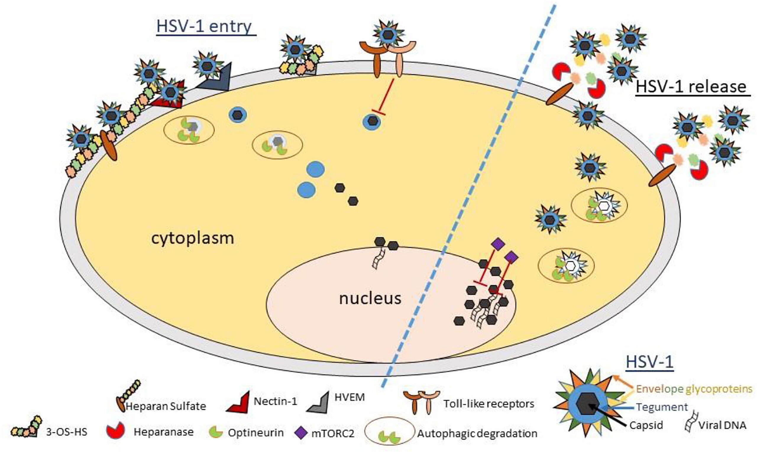
English: Figure 1. Schematic diagram and model showing important host proteins and their roles in pathophysiology of ocular herpes infection. HSV-1 entry into a corneal cell starts with the binding of viral glycoproteins gB and/or gC to cell surface heparan sulfate. Next, HSV-1 gD binds to one of its cognate receptors (Nectin-1, HVEM, and 3-OS HS) to start viral capsid penetration into the cytoplasm. Toll-like receptors sense viral invasion and activate intrinsic immune responses to control infection. A newly identified restriction factor, optineurin, reduces incoming or outgoing viral load by selective degradation of viral capsid and essential proteins via autophagy. In parallel, mTORC2 complex acts to reduce viral replication in the nucleus. In a pro-viral role, heparanase, a host enzyme is upregulated to facilitate HSV-1 release from the corneal cell by removal of cell surface HS. While the events summarized here do not describe the complete picture of viral invasion, they do highlight many new findings and interventional targets in context with HSV-1 infection of the cornea. A cartoon describing the essential structural components of a matured HSV-1 virion is shown (bottom right). |
| Date | |
| Source | https://www.frontiersin.org/journals/microbiology/articles/10.3389/fmicb.2022.818658/full |
| Author | Sajal Deea Shukla Tibor Valyi-Nagy |
Licensing
[edit]
This file is licensed under the Creative Commons Attribution 4.0 International license.
- You are free:
- to share – to copy, distribute and transmit the work
- to remix – to adapt the work
- Under the following conditions:
- attribution – You must give appropriate credit, provide a link to the license, and indicate if changes were made. You may do so in any reasonable manner, but not in any way that suggests the licensor endorses you or your use.

|
This media file is uncategorized.
Please help improve this media file by adding it to one or more categories, so it may be associated with related media files (how?), and so that it can be more easily found.
Please notify the uploader with {{subst:Please link images|File:Fmicb-13-818658-g001.jpg}} ~~~~ |
File history
Click on a date/time to view the file as it appeared at that time.
| Date/Time | Thumbnail | Dimensions | User | Comment | |
|---|---|---|---|---|---|
| current | 21:40, 13 May 2024 | | 4,317 × 2,583 (540 KB) | Ozzie10aaaa (talk | contribs) | Uploaded a work by Sajal Deea Shukla Tibor Valyi-Nagy from https://www.frontiersin.org/journals/microbiology/articles/10.3389/fmicb.2022.818658/full with UploadWizard |
You cannot overwrite this file.
File usage on Commons
There are no pages that use this file.