File:Frontal histological sections of Zoea IV (larval stage of the European shore crab Carcinus maenas).png
Jump to navigation
Jump to search

Size of this preview: 455 × 600 pixels. Other resolutions: 182 × 240 pixels | 364 × 480 pixels | 582 × 768 pixels | 777 × 1,024 pixels | 1,882 × 2,481 pixels.
Original file (1,882 × 2,481 pixels, file size: 6.7 MB, MIME type: image/png)
Captions
Captions
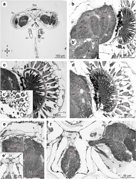
Frontal histological sections of Zoea IV (larval stage of the European shore crab Carcinus maenas), showing anterior part of the head with stalked compound eyes and eyestalk neuropils.
Summary[edit]
.png&action=edit§ion=1)
| Description |
English: Frontal histological sections of Zoea IV (Holländer stain). a - overview of the anterior part of the head with stalked compound eyes and eyestalk neuropils (EyNP), see Fig. Fig.2c2c for section plane; b - higher magnification of the right compound eye showing the visual neuropils lamina (La), medulla (Me), and lobula (Lo), proximally adjoined by the complex of the hemiellipsoid body neuropil and the medulla terminalis (HN/TM); b` - visual chiasm (arrow head) between medulla and lamina; c – the compound eye consists of many single ommatidia (one ommatidium surrounded by a line), note that the cornea is detached because of poor fixation (↔ double arrow); c`- cross sections of ommatidia show that the crystalline cones are composed of four cone cells. Each cell of one crystalline cone is indicated by an arrowhead; d - the ommatidia extend bundles of neurites (Neu) into the lamina; e – a branch of the ophthalmic artery (OA) extends towards the eyestalk neuropils; e`- the two main branches of the ophthalmic artery (OA) course towards the eyestalks; f – the protocerebral tract (PCT) connects the eyestalk neuropils and protocerebrum (PC) of median brain. Abbreviations: A1 - first antenna, Cap - capillary, CL - corneal lens, Co - cornea of compound eye, Cry - crystalline cone, Cu - cuticle, DP - distal pigment, Ey - compound eye, EyNP - eyestalk neuropils, HN/TM - complex of hemiellipsoid body neuropil and medulla terminalis, La - lamina, Lo - lobula, Me - medulla, Neu - neurites, OA - ophthalmic artery, Om - ommatidium of compound eye, PC - protocerebrum, PCT - protocerebral tract, Pi - shielding pigment, Rh - rhabdomeres of photoreceptor cell, Ros - rostrum, RS - rostral spine.
Spitnzer et al., 2018 (https://dx.doi.org/10.1186/s12983-018-0271-z) |
| Date | |
| Source | https://doi.org/10.1186/s12983-018-0271-z |
| Author | Franziska Spitzner, Rebecca Meth,Christina Krüger, Emanuel Nischik, Stefan Eiler, Andy Sombke, Gabriela Torres, Steffen Harzsch |
Licensing[edit]
.png&action=edit§ion=2)
This file is licensed under the Creative Commons Attribution 4.0 International license.
- You are free:
- to share – to copy, distribute and transmit the work
- to remix – to adapt the work
- Under the following conditions:
- attribution – You must give appropriate credit, provide a link to the license, and indicate if changes were made. You may do so in any reasonable manner, but not in any way that suggests the licensor endorses you or your use.
File history
Click on a date/time to view the file as it appeared at that time.
| Date/Time | Thumbnail | Dimensions | User | Comment | |
|---|---|---|---|---|---|
| current | 11:05, 22 August 2021 | | 1,882 × 2,481 (6.7 MB) | Dom (UZH) (talk | contribs) | Uploaded a work by Franziska Spitzner, Rebecca Meth,Christina Krüger, Emanuel Nischik, Stefan Eiler, Andy Sombke, Gabriela Torres, Steffen Harzsch from https://doi.org/10.1186/s12983-018-0271-z with UploadWizard |
You cannot overwrite this file.
File usage on Commons
There are no pages that use this file.
File usage on other wikis
The following other wikis use this file:
- Usage on en.wikibooks.org
.png&oldid=657423683)