File:Gray219.png
Originaldatei (650 × 831 Pixel, Dateigröße: 76 KB, MIME-Typ: image/png)
Bildtexte
Kurzbeschreibungen
Beschreibung
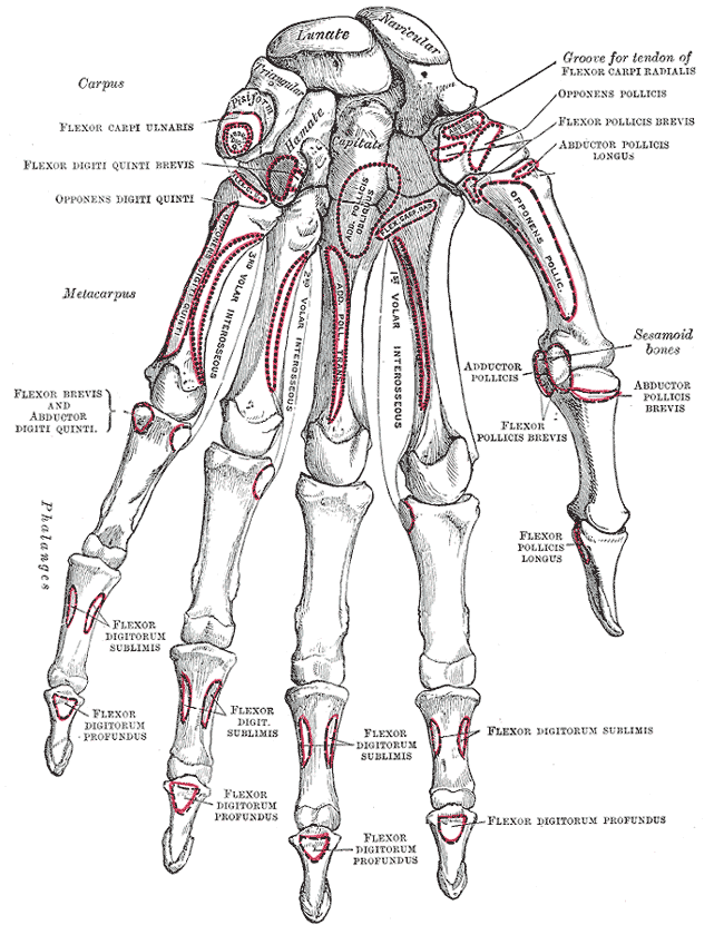
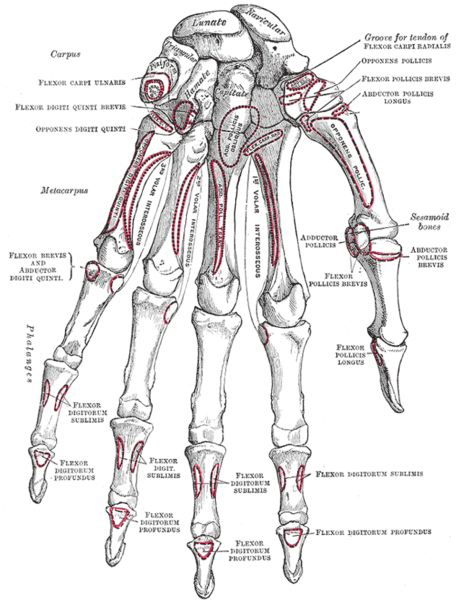
[Bearbeiten]| Beschreibung | Bones of the left hand. Volar surface. | ||||||||||||||||||||
| Tafel | 219 | ||||||||||||||||||||
| Datum | vor 1858 | ||||||||||||||||||||
| Quelle |
|
||||||||||||||||||||
| Urheber |
|
||||||||||||||||||||
.jpg)
Buch
[Bearbeiten]| Henry Gray: Gray's Anatomy. - 20. Aufl. 1918
|
|||||||||||||||||||||||
|---|---|---|---|---|---|---|---|---|---|---|---|---|---|---|---|---|---|---|---|---|---|---|---|
| Urheber |
|
-_Title_page.png) | |||||||||||||||||||||
| Herausgeber |
Revised by Warren H. Lewis |
||||||||||||||||||||||
| Illustrator |
|
||||||||||||||||||||||
| Titel | |||||||||||||||||||||||
| Auflage |
20 |
||||||||||||||||||||||
| Verleger | |||||||||||||||||||||||
| Objektart |
Ausgabe oder Version |
||||||||||||||||||||||
| Seitenübersicht | list of all the plates | ||||||||||||||||||||||
| Sprache |
Englisch |
||||||||||||||||||||||
| Veröffentlichungsdatum |
1918 |
||||||||||||||||||||||
| Erscheinungsort |
Philadelphia / New York |
||||||||||||||||||||||
| Quelle | Bartleby | ||||||||||||||||||||||

Lizenz
[Bearbeiten]
Dieses Bild ist gemeinfrei, weil es ein rein mechanischer Scan oder eine rein mechanische Fotografie eines gemeinfreien Originals ist, oder – wenn anders belegbar – es einem solchen Scan oder einer solchen Fotografie so ähnlich ist, dass das Entstehen eines Copyright-Schutzes nicht erwartet werden kann. Das Original selbst ist gemeinfrei aus folgendem Grund:
Dieser Hinweis wurde für Fälle entworfen, in denen erklärt werden muss, dass jegliche Verbesserungen (zum Beispiel von Helligkeit, Kontrast, Farbabgleich, Schärfe) für sich selbst nicht ausreichend sind, um ein neues Copyright zu schaffen. Er kann verwendet werden, wenn unbekannt ist, ob Verbesserungen vorgenommen wurden, oder wenn Verbesserungen erkennbar, aber nicht ausreichend sind. Für bekanntermaßen unbearbeitete Scans kannst Du statt dessen den passenden {{PD-old}}-Hinweis verwenden. Anwendungshinweise findest Du unter Commons:When to use the PD-scan tag.  | ||||
Dateiversionen
Klicke auf einen Zeitpunkt, um diese Version zu laden.
| Version vom | Vorschaubild | Maße | Benutzer | Kommentar | |
|---|---|---|---|---|---|
| aktuell | 22:18, 17. Mär. 2007 | | 650 × 831 (76 KB) | Tene~commonswiki (Diskussion | Beiträge) | Optimised (0) |
| 18:21, 13. Apr. 2005 |  | 650 × 831 (78 KB) | Phyzome (Diskussion | Beiträge) | {{Gray's Anatomy plate| Bones of the left hand. Volar surface.}} |
Du kannst diese Datei nicht überschreiben.
Dateiverwendung
Mehr als 100 Seiten verwenden diese Datei. Die folgende Liste zeigt nur die ersten 100 Verwendungen dieser Datei. Eine vollständige Liste ist verfügbar.
- Bones
- Schemes/en/Human anatomy
- File:Anaphase.jpg
- File:Augennerven.jpg
- File:Biceps femoris muscle long head.PNG
- File:Biceps femoris muscle short head.PNG
- File:Brain diagram ja.png
- File:Brain diagram ja.svg
- File:Bulbospongiosus-Female.png
- File:Bulbospongiosus-Male.png
- File:Circulus arteriosus SAB Lokalisationen.png
- File:Coracobrachialis.png
- File:ECR-brevis.png
- File:Extensor carpi radialis longus.png
- File:Extensor digitorum longus.png
- File:Extensor hallucis longus.png
- File:Eyemuscles.png
- File:Femur head.png
- File:Gallenblase.png
- File:Gastro-intestinal tract.png
- File:Gemellus superior muscle.PNG
- File:Glosso.gif
- File:Gluteus maximus muscle.PNG
- File:Gluteus medius muscle.PNG
- File:Gluteus minimus muscle.PNG
- File:Gluteus muscles.PNG
- File:Gray219.png
- File:Hand bone.png
- File:Heart-and-lungs.jpg
- File:Histologie der Nebenniere.png
- File:Hypoglossus.jpg
- File:Inferior gemellus muscle.PNG
- File:Ischiadicus.jpg
- File:Ischiocavernosus-female.png
- File:Ischiocavernosus-male.png
- File:Kidney nephron.png
- File:Levator ani.png
- File:Longus capitis.png
- File:Longus colli.png
- File:Lungs anatomy.png
- File:Mandibularis.gif
- File:Musculus abductor digiti minimi (Hand).png
- File:Musculus abductor pollicis brevis.png
- File:Musculus cremaster.png
- File:Musculus flexor pollicis longus.png
- File:Musculus frontalis.png
- File:Musculus occipitalis.png
- File:Musculus omohyoideus.png
- File:Nasennebenhöhlen.gif
- File:Nervusfemoralis.jpg
- File:Nuchal lines.png
- File:Operculum.png
- File:Orrsövény.PNG
- File:Otosklerose.png
- File:Pankreas - Topografie.png
- File:Pectineal line.png
- File:Pectineus.png
- File:Peroneus tertius.png
- File:Peroneusbrevis.png
- File:Petrotympanic fissure.png
- File:Piriformis muscle.PNG
- File:Platysma.png
- File:Plexuslumbosacralis.jpg
- File:Prepatellar bursa.png
- File:Pronator-quadratus.png
- File:Pronator-teres.png
- File:Quadratus femoris muscle.PNG
- File:Rectus capitis lateralis muscle.PNG
- File:Rectus femoris.png
- File:Sartorius muscle.png
- File:Scalenus anterior.png
- File:Scalenus medius.png
- File:Scalenus posterior.png
- File:Schaedel-mensch-seitenansicht.jpg
- File:Semimembranosus muscle.PNG
- File:Semitendinosus muscle.PNG
- File:Situs inversus - Mirrored heart and lungs.jpg
- File:Spine of sphenoid bone.png
- File:Sternohyoideus.png
- File:Sternothyroideus.png
- File:Stylomastoid foramen.png
- File:Surface anatomy of the back-Gray.png
- File:Telencephalon-Horiconatal.jpg
- File:Tensor fasciae latae.png
- File:Thigh muscles back.png
- File:Thymus.png
- File:Thymus de.png
- File:Thyrohyoideus.png
- File:Tibialis anterior 2.png
- File:Tibialis posterior.png
- File:Tongue-sour.jpg
- File:Tongue-sweet.jpg
- File:Transversus abdominis.png
- File:Transversus thoracis.png
- File:Tuberosity of the ischium.PNG
- File:Vaginaschnitt.gif
- File:Vagusgruppe Ganglien.jpg
- File:Vastus lateralis muscle.png
- File:Vastus medialis muscle.png
- File:WeiblicherGenitaltrakt.gif
.png)
Weitere Links auf diese Datei.
Globale Dateiverwendung
Die nachfolgenden anderen Wikis verwenden diese Datei:
- Verwendung auf ar.wikipedia.org
- Verwendung auf arz.wikipedia.org
- Verwendung auf ast.wikipedia.org
- Verwendung auf as.wikipedia.org
- Verwendung auf az.wikipedia.org
- Verwendung auf bg.wikipedia.org
- Verwendung auf bn.wikipedia.org
- Verwendung auf br.wikipedia.org
- Verwendung auf bs.wikipedia.org
- Verwendung auf ca.wikipedia.org
- Múscul flexor comú profund dels dits
- Múscul flexor llarg del polze
- Anatomia de Gray
- Carp
- Metacarp
- Escafoide del carp
- Os piramidal
- Ganxut
- Trapezoide (os)
- Trapezi (os)
- Gran del carp
- Pisiforme
- Semilunar
- Múscul cubital anterior
- Múscul flexor comú superficial dels dits
- Múscul oponent del polze
- Múscul flexor curt del polze
- Múscul oponent del menovell
- Múscul flexor curt del menovell
- Verwendung auf cv.wikipedia.org
- Verwendung auf cy.wikipedia.org
- Verwendung auf da.wikipedia.org
- Verwendung auf de.wikibooks.org
- Verwendung auf en.wikipedia.org
- Gray's Anatomy
- Flexor carpi ulnaris muscle
- Flexor pollicis brevis muscle
- Flexor pollicis longus muscle
- Opponens digiti minimi muscle of hand
- Opponens pollicis muscle
- Flexor digiti minimi brevis muscle of hand
- Talk:Abductor pollicis longus muscle
- Hand
- Talk:Extrinsic extensor muscles of the hand
- User:Was a bee/Gray
- Verwendung auf eo.wikipedia.org
- Verwendung auf es.wikipedia.org
Weitere globale Verwendungen dieser Datei anschauen.
- Gray's Anatomy plates of bones
- Anatomical plates and drawings of the bones of the human hand
- Human metacarpus
- Trapezium (bone)
- Trapezoid bone
- Pisiform bone
- Triquetral bone
- Scaphoid bone
- Capitate bone
- Hamate bone
- Lunate bone
- Human hand bone with muscle attachments
- Human metacarpus with muscle attachments
- Human pisiform bone with muscle attachments
- Human hamate bone with muscle attachments
- Human phalanges of hand with muscle attachments
- Human carpal bones with muscle attachments