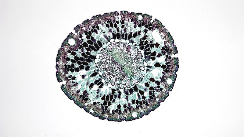
File:Gymnosperm Leaves Single Needled Pinus (36445773426).jpg

Original file (3,264 × 1,840 pixels, file size: 3.01 MB, MIME type: image/jpeg)
Captions
Captions
Summary[edit]
| DescriptionGymnosperm Leaves Single Needled Pinus (36445773426).jpg |
cross section: Pinus needle magnification: 40x Pinus needles may be circular, semicircular or triangular in cross section and are structurally adapted to tolerate xeric stress and freezing. A single layered epidermis of lignified cells is covered by a heavy waxy cuticle. Numerous sunken stomata are scattered over the entire epidermal surface. They are bounded by pairs of subsidiary cells, and open internally to a substomatal cavity and externally to a respiratory cavity or vestibule. The underlying hypodermis consists of several layers of thick-walled sclerenchyma, particularly well-developed at ridges. Deep to the hypodermis lies an undifferentiated photosynthetic mesophyll. Cells are distinctively lobed with infolded cell walls. They are filled with chloroplasts and starch grains that may be difficult to see because of an accumulation of dark staining tannins and resins. A few resin canals can be seen close to the hypodermal side of the mesophyll. A central vascular bundle is wrapped in a single layered endodermis with a well-defined casparian strip whose end walls may become heavily suberized with age. Deep to the endodermis lies a multilayered parenchymous pericycle with two vascular bundles separated by an often indistinct band of sclerenchymal cells. The pericycle also contains transfusion tissues of protein rich albuminous cells which abut and assist the phloem in the transport nutrients and elongated tracheidal cells which abut and assists the xylem in the transport of water. Most species possess two conjoint, collateral vascular bundles, surrounded and supported by the tissues of the pericycle. Xylem tracheids lie towards the adaxial surface and phloem sieve tubes towards abaxial surface of the leaf. Xylem vessels and fibers, and phloem companion cells are absent in Gymnosperms. While vascular bundles are closed some cambium may persist near the base of the needle. Visit the BCC Bioscience Image featuring the Microscopic World of Plants. <a href="http://www.berkshirecc.edu/biologyimages" rel="nofollow">www.berkshirecc.edu/biologyimages</a> |
| Date | |
| Source | Gymnosperm Leaves: Single Needled Pinus |
| Author | Berkshire Community College Bioscience Image Library |
Licensing[edit]
| This file is made available under the Creative Commons CC0 1.0 Universal Public Domain Dedication. | |
| The person who associated a work with this deed has dedicated the work to the public domain by waiving all of their rights to the work worldwide under copyright law, including all related and neighboring rights, to the extent allowed by law. You can copy, modify, distribute and perform the work, even for commercial purposes, all without asking permission.
http://creativecommons.org/publicdomain/zero/1.0/deed.enCC0Creative Commons Zero, Public Domain Dedicationfalsefalse |
| This image was originally posted to Flickr by bccoer at https://flickr.com/photos/146824358@N03/36445773426 (archive). It was reviewed on 22 June 2018 by FlickreviewR 2 and was confirmed to be licensed under the terms of the cc-zero. |
22 June 2018
File history
Click on a date/time to view the file as it appeared at that time.
| Date/Time | Thumbnail | Dimensions | User | Comment | |
|---|---|---|---|---|---|
| current | 23:55, 22 June 2018 |  | 3,264 × 1,840 (3.01 MB) | Meisam (talk | contribs) | Transferred from Flickr via #flickr2commons |
You cannot overwrite this file.
File usage on Commons
There are no pages that use this file.
Metadata
This file contains additional information such as Exif metadata which may have been added by the digital camera, scanner, or software program used to create or digitize it. If the file has been modified from its original state, some details such as the timestamp may not fully reflect those of the original file. The timestamp is only as accurate as the clock in the camera, and it may be completely wrong.
| Date and time of data generation | 11:00, 10 February 2014 |
|---|---|
| Horizontal resolution | 1 dpc |
| Vertical resolution | 1 dpc |
| Software used | Windows Photo Editor 10.0.10011.16384 |
| File change date and time | 21:21, 10 August 2017 |
| Y and C positioning | Centered |
| Exif version | 2.3 |
| Date and time of digitizing | 11:00, 10 February 2014 |
| Meaning of each component |
|
| DateTimeOriginal subseconds | 00 |
| DateTimeDigitized subseconds | 00 |
| Supported Flashpix version | 1 |
| Color space | sRGB |