File:In vivo AO 2PFM visualized features of epithelium and fiber cell organization in the mouse lens.jpg

Original file (682 × 684 pixels, file size: 190 KB, MIME type: image/jpeg)
Captions
Captions
Summary
[edit]
| Description |
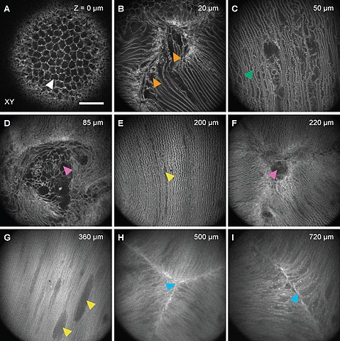
English: In vivo AO 2PFM visualizes features of epithelium and fiber cell organization in mouse lenses.
(A-I) Representative images of cellular features at various depths visualized by AO 2PFM in the anterior lenses of the mice employed in the study. (A) Lens epithelium, (B) disorganized and broadened fiber cell ends, (C) non-uniform superficial fiber cells with excessive interdigitation, (D, F) anterior voids, (E, G) mostly uniform rows of fiber cells with enlarged vacuoles, (H, I) tight suture structures at depth. Depth noted in each panel is relative to the epithelium. Arrowheads point to features described in the main text. Scale bar: 50 μm. |
| Date | |
| Source |
Version 1. bioRxiv. Preprint. 2023 Jan 19. doi: 10.1101/2023.01.17.524320 PMCID: PMC9882239 PMID: 36711806 |
| Author |
Santosh Kumar Paidi,1 Qinrong Zhang,2,3 Yuhan Yang,2 Chun-Hong Xia,1,4 Na Ji,2,3,5,6,7 and Xiaohua Gong1,4,7 1School of Optometry, University of California, Berkeley, California 94720, USA 2Department of Physics, University of California, Berkeley, California 94720, USA 3Department of Molecular and Cell Biology, University of California, Berkeley, CA 94720, USA 4Vision Science Program, University of California, Berkeley, California 94720, USA 5Helen Wills Neuroscience Institute, University of California, Berkeley, CA 94720, USA 6Molecular Biophysics and Integrated Bioimaging Division, Lawrence Berkeley National Laboratory, Berkeley, CA 94720, USA |
Licensing
[edit]
- You are free:
- to share – to copy, distribute and transmit the work
- to remix – to adapt the work
- Under the following conditions:
- attribution – You must give appropriate credit, provide a link to the license, and indicate if changes were made. You may do so in any reasonable manner, but not in any way that suggests the licensor endorses you or your use.
- share alike – If you remix, transform, or build upon the material, you must distribute your contributions under the same or compatible license as the original.
File history
Click on a date/time to view the file as it appeared at that time.
| Date/Time | Thumbnail | Dimensions | User | Comment | |
|---|---|---|---|---|---|
| current | 20:38, 13 February 2023 | | 682 × 684 (190 KB) | Tgru001 (talk | contribs) | Uploaded a work by Santosh Kumar Paidi,1 Qinrong Zhang,2,3 Yuhan Yang,2 Chun-Hong Xia,1,4 Na Ji,2,3,5,6,7 and Xiaohua Gong1,4,7 1School of Optometry, University of California, Berkeley, California 94720, USA 2Department of Physics, University of California, Berkeley, California 94720, USA 3Department of Molecular and Cell Biology, University of California, Berkeley, CA 94720, USA 4Vision Science Program, University of California, Berkeley, California 94720, USA 5Helen Wills Neuroscience Insti... |
You cannot overwrite this file.
File usage on Commons
There are no pages that use this file.
File usage on other wikis
The following other wikis use this file:
- Usage on en.wikipedia.org