File:Inclusion body myositis histology.jpg
Jump to navigation
Jump to search
Size of this preview: 534 × 600 pixels. Other resolutions: 214 × 240 pixels | 536 × 602 pixels.
Original file (536 × 602 pixels, file size: 123 KB, MIME type: image/jpeg)
Captions
Captions
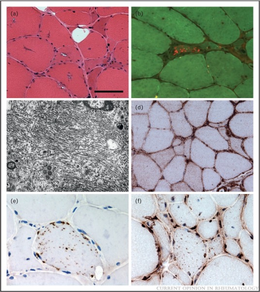
Microscopic cross sections of muscle in sIBM with various stains
Summary[edit]
| Description |
English: "Pathological features observed in IBM. Muscle biopsy from a patient with IBM showing fibres containing rimmed vacuoles (a), amyloid in a tissue section stained using Congo red and visualised under fluorescent light (b) and tubulofilaments observed using electron microscopy (c). Immunohistochemically stained tissue sections reveal increased sarcolemmal and sarcoplasmic major histocompatibility complex class I (MHC Class I) expression (d) and fibres containing sarcoplasmic p62 immunoreactive aggregates (e) and TAR DNA-binding protein 43 (TDP-43) immunoreactive aggregates with loss of normal myonuclear TDP-43 staining (f). Scale bar in A represents 50 μm in (a), (b), (d), (f); 25 μm in (e); and 0.7 μm in (c)."
Copied from caption of cited article. |
| Date | |
| Source | Update in inclusion body myositis. Current Opinion in Rheumatology: November 2013 - Volume 25 - Issue 6 - p 763-771 doi: 10.1097/01.bor.0000434671.77891.9a |
| Author | Machado, Pedro*; Brady, Stefen*; Hanna, Michael G. |
Licensing[edit]
This file is licensed under the Creative Commons Attribution 3.0 Unported license.
- You are free:
- to share – to copy, distribute and transmit the work
- to remix – to adapt the work
- Under the following conditions:
- attribution – You must give appropriate credit, provide a link to the license, and indicate if changes were made. You may do so in any reasonable manner, but not in any way that suggests the licensor endorses you or your use.
File history
Click on a date/time to view the file as it appeared at that time.
| Date/Time | Thumbnail | Dimensions | User | Comment | |
|---|---|---|---|---|---|
| current | 00:13, 1 May 2022 | | 536 × 602 (123 KB) | Lukelahood (talk | contribs) | Uploaded a work by Machado, Pedro*; Brady, Stefen*; Hanna, Michael G. from Update in inclusion body myositis. Current Opinion in Rheumatology: November 2013 - Volume 25 - Issue 6 - p 763-771 doi: 10.1097/01.bor.0000434671.77891.9a with UploadWizard |
You cannot overwrite this file.
File usage on Commons
There are no pages that use this file.
File usage on other wikis
The following other wikis use this file:
- Usage on en.wikipedia.org