File:Invasive Breast Cancer in 3D.jpg
Jump to navigation
Jump to search
Size of this preview: 720 × 599 pixels. Other resolutions: 288 × 240 pixels | 577 × 480 pixels | 923 × 768 pixels | 1,230 × 1,024 pixels | 1,557 × 1,296 pixels.
Original file (1,557 × 1,296 pixels, file size: 597 KB, MIME type: image/jpeg)
Captions
Captions
Add a one-line explanation of what this file represents
Summary
[edit]
| Description |
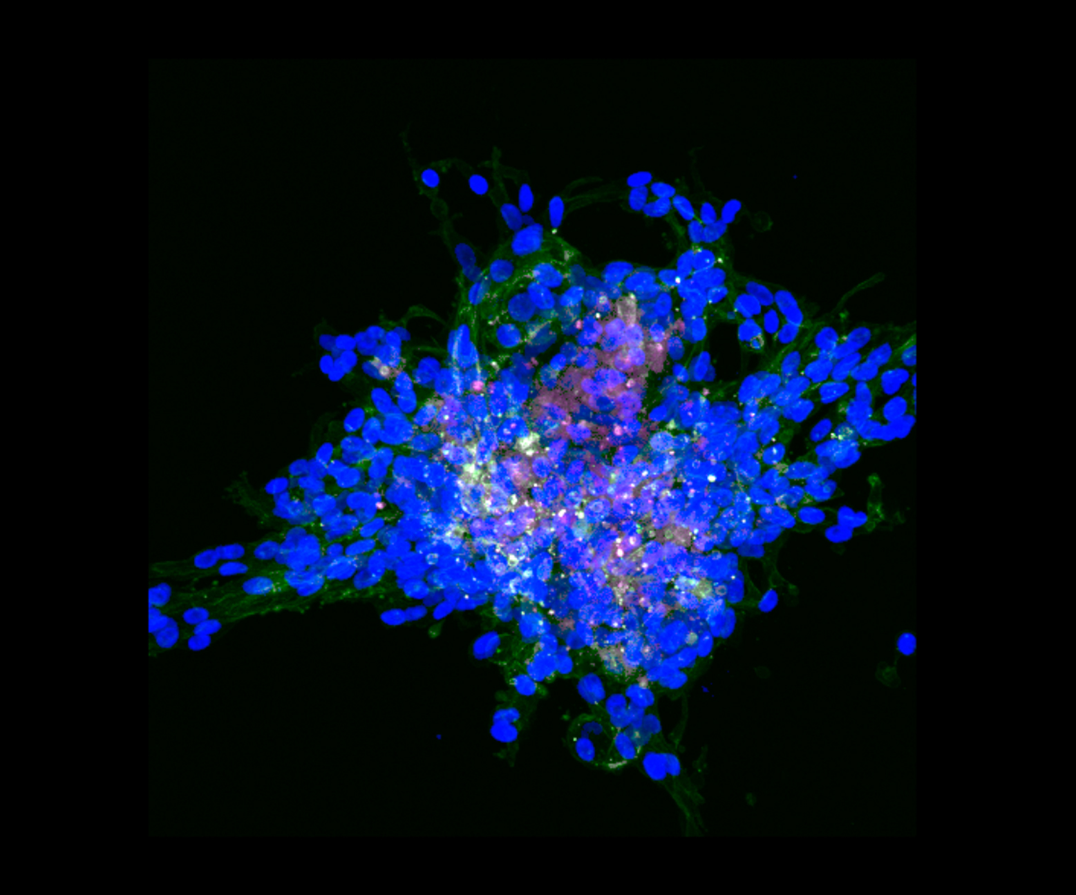
English: This is an invasive breast tumor, grown from invasive breast cancer cells in a matrix that mimics human breast tissue. The blue is a stain of the nucleus of the cell. The green is F-actin, which contributes to the structure and shape of the cell. The white/pink in the middle is Vimentin, which also contributes to the structure. Notice how the cells in the middle of the tumor have the most Vimentin. These might give these cells the ability to “lead the pack” and metastasize to other areas.
Other Information: The image is a collapsed reconstruction of 58 z-stacks taken of the tumor, taken at 20x magnification with a Zeiss LSM 780 fluorescent microscope. Taken at Wayne State University, Detroit, MI. |
| Date | |
| Source | Own work |
| Author | Sshah74 |
Licensing
[edit]
I, the copyright holder of this work, hereby publish it under the following license:
This file is licensed under the Creative Commons Attribution 4.0 International license.
- You are free:
- to share – to copy, distribute and transmit the work
- to remix – to adapt the work
- Under the following conditions:
- attribution – You must give appropriate credit, provide a link to the license, and indicate if changes were made. You may do so in any reasonable manner, but not in any way that suggests the licensor endorses you or your use.
| This image was uploaded as part of Wiki Science Competition 2019. |
File history
Click on a date/time to view the file as it appeared at that time.
| Date/Time | Thumbnail | Dimensions | User | Comment | |
|---|---|---|---|---|---|
| current | 21:07, 12 December 2019 | | 1,557 × 1,296 (597 KB) | Sshah74 (talk | contribs) | User created page with UploadWizard |
You cannot overwrite this file.
File usage on Commons
There are no pages that use this file.