File:Journal.ppat.1005203.g001.D.jpg
Jump to navigation
Jump to search
Size of this preview: 800 × 430 pixels. Other resolutions: 320 × 172 pixels | 640 × 344 pixels | 1,024 × 550 pixels | 1,280 × 688 pixels | 1,980 × 1,064 pixels.
Original file (1,980 × 1,064 pixels, file size: 2.1 MB, MIME type: image/jpeg)
Captions
Captions
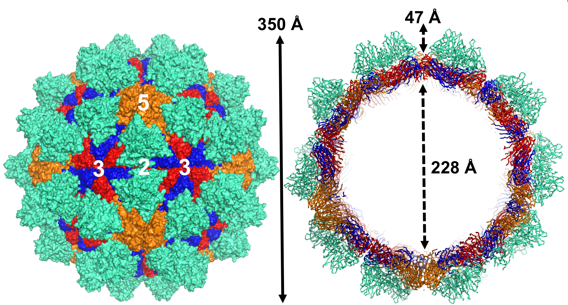
The overall structure of grouper nervous necrosis virus (GNNV).
Summary[edit]
| Description |
English: The overall structure of grouper nervous necrosis virus (GNNV). Surface domain-colored diagram (left) and central cavity (right) representations of the T = 3 GNNV-LP. The tip-to-tip distance is ~350 Å, the diameter of the central cavity is ~228 Å, and the spike protrusion on the capsid surface is ~47 Å. The S-domains of the subunits A, B and C are shown in orange, blue and red, respectively, and the P-domains are shown in cyan. The structure of the GNNV-LP is viewed along the I2, I3 and I5 axes. |
| Date | |
| Source | Chen N-C, Yoshimura M, Guan H-H, Wang T-Y, Misumi Y, Lin C-C, et al. (2015) Crystal Structures of a Piscine Betanodavirus: Mechanisms of Capsid Assembly and Viral Infection. PLoS Pathog 11(10): e1005203. https://doi.org/10.1371/journal.ppat.1005203 |
| Author | Nai-Chi Chen, Masato Yoshimura, Hong-Hsiang Guan, Ting-Yu Wang, Yuko Misumi, Chien-Chih Lin, Phimonphan Chuankhayan, Atsushi Nakagawa, Sunney I. Chan, Tomitake Tsukihara, Tzong-Yueh Chen, Chun-Jung Chen |
| Other versions |
  |
Licensing[edit]
This file is licensed under the Creative Commons Attribution 4.0 International license.
- You are free:
- to share – to copy, distribute and transmit the work
- to remix – to adapt the work
- Under the following conditions:
- attribution – You must give appropriate credit, provide a link to the license, and indicate if changes were made. You may do so in any reasonable manner, but not in any way that suggests the licensor endorses you or your use.
File history
Click on a date/time to view the file as it appeared at that time.
| Date/Time | Thumbnail | Dimensions | User | Comment | |
|---|---|---|---|---|---|
| current | 11:58, 6 December 2020 | | 1,980 × 1,064 (2.1 MB) | Guest2625 (talk | contribs) | Uploaded a work by Nai-Chi Chen, Masato Yoshimura, Hong-Hsiang Guan, Ting-Yu Wang, Yuko Misumi, Chien-Chih Lin, Phimonphan Chuankhayan, Atsushi Nakagawa, Sunney I. Chan, Tomitake Tsukihara, Tzong-Yueh Chen, Chun-Jung Chen from Chen N-C, Yoshimura M, Guan H-H, Wang T-Y, Misumi Y, Lin C-C, et al. (2015) Crystal Structures of a Piscine Betanodavirus: Mechanisms of Capsid Assembly and Viral Infection. PLoS Pathog 11(10): e1005203. https://doi.org/10.1371/journal.ppat.1005203 with UploadWizard |
You cannot overwrite this file.
File usage on Commons
The following 2 pages use this file:
File usage on other wikis
The following other wikis use this file:
- Usage on de.wikipedia.org
- Usage on en.wikipedia.org
- Usage on uk.wikipedia.org
- Usage on zh.wikipedia.org