File:Kidney tubules.png
跳转到导航
跳转到搜索
没有更高的分辨率。
Kidney_tubules.png (256 × 256像素,文件大小:118 KB,MIME类型:image/png)
说明
说明
添加一行文字以描述该文件所表现的内容
摘要
[编辑]
| 描述 |
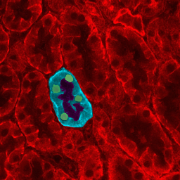
English: Microscopy of kidney tissue showing tubules. One tubule is highlighted to show epithelial cells (blue), cell nuclei (green) and the tubule lumen (dark center). Source: my personal image. The copyright to this image is retained by John Schmidt (JWSchmidt). Permission is granted to copy, distribute and/or modify this image under the terms of the Wikipedia GFDL, as indicated in the fine print at the bottom of this page. If you do not want to use this image under the terms of the GFDL, you can alternatively use it under the terms of the cc-by-nc-sa license.
Türkçe: Böbrek dokusu, tübüller gözükmekte. Tübüllerden biri epitel hücreleri (mavi), hücre çekirdeklerini (yeşil) ve tübül deliğini (koyu merkez/orta) göstermek için ışıklandırılmıştır. |
| 日期 | 2004年4月2日 (原始上传日期) |
| 来源 | Transferred from en.wikipedia |
| 作者 | Original uploader was JWSchmidt at en.wikipedia |
| 授权 (二次使用本文件) |
GFDL-SELF-WITH-DISCLAIMERS; Released under the GNU Free Documentation License. |
许可协议
[编辑]

|
已授权您依据自由软件基金会发行的无固定段落及封面封底文字(Invariant Sections, Front-Cover Texts, and Back-Cover Texts)的GNU自由文件许可协议1.2版或任意后续版本的条款,复制、传播和/或修改本文件。该协议的副本请见“GNU Free Documentation License”。 |
| 本文件采用知识共享署名-相同方式共享 3.0 未本地化版本许可协议授权。 | ||
| ||
| 本许可协议标签作为GFDL许可协议更新的组成部分被添加至本文件。 |
我,本作品著作权人,特此采用以下许可协议发表本作品:

|
已授权您依据自由软件基金会发行的无固定段落及封面封底文字(Invariant Sections, Front-Cover Texts, and Back-Cover Texts)的GNU自由文件许可协议1.2版或任意后续版本的条款,复制、传播和/或修改本文件。该协议的副本请见“GNU Free Documentation License”。 受免責聲明的約束。 |
原始上传日志
[编辑]
The original description page was here. All following user names refer to en.wikipedia.
- 2004-04-02 19:29 JWSchmidt 256×256×8 (121209 bytes) Microscopy of kidney tissue showing tubules.
文件历史
点击某个日期/时间查看对应时刻的文件。
| 日期/时间 | 缩略图 | 大小 | 用户 | 备注 | |
|---|---|---|---|---|---|
| 当前 | 2008年1月8日 (二) 15:10 | | 256 × 256(118 KB) | Maderibeyza(留言 | 贡献) | {{Information |Description={{en|Microscopy of kidney tissue showing tubules. One tubule is highlighted to show epithelial cells (blue), cell nuclei (green) and the tubule lumen (dark center). Source: my personal image. The copyright to this image is retai |
您不可以覆盖此文件。
文件用途
以下页面使用本文件:
全域文件用途
以下其他wiki使用此文件:
- ar.wikipedia.org上的用途
- bs.wikipedia.org上的用途
- en.wikipedia.org上的用途
- fa.wikipedia.org上的用途
- id.wikipedia.org上的用途
- sr.wikipedia.org上的用途
- ur.wikipedia.org上的用途
- zh.wikipedia.org上的用途