File:LDs are distributed cortically in the zebrafish blastodisc.jpg
Original file (1,451 × 1,024 pixels, file size: 747 KB, MIME type: image/jpeg)
Captions
Captions
Summary[edit]
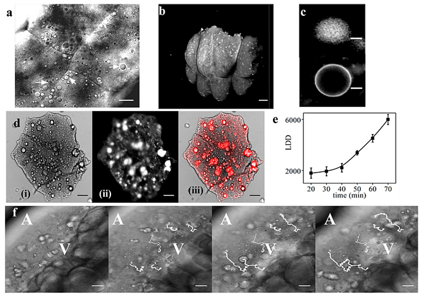
| Description | Figure 1. Lipid droplets (LDs) are distributed cortically in the zebrafish blastodisc.: (a) DIC image of zebrafish blastodisc at 4 cell stage (animal-pole view). Arrowheads point to the LDs. (b) Nile Red labeled 3D-rendered confocal image of blastodisc showing cortical distribution of LDs. (c) Nile Red stained images of LD (upper panel) and artificially synthesized giant vesicle (lower panel). (d) (i) DIC (ii) fluorescent (iii) merge images, of Nile Red stained de-yolked blastodisc showing co-localization of Nile Red signal (ii) with the granules of (i) conforming LD identity. (e) Mean LDD versus time averaged over three embryos, error bars are standard error of mean (SEM). (f) Time lapse images of blastodisc (lateral orientation) showing migration of LDs to cortical regions of the embryo. The white lines denote the trajectories of 5 representative LDs. ‘A’ and ‘V’ denote the animal and vegetal pole of the embryo. Scale bar 25 μm in (a) and (f), 8 μm in (c), 150 μm in (b) and 100 μm in (d). |
| Date | |
| Source |
https://www.researchgate.net/publication/281676795_Turnover_of_the_actomyosin_complex_in_zebrafish_embryos_directs_geometric_remodelling_and_the_recruitment_of_lipid_droplets Turnover of the actomyosin complex in zebrafish embryos directs geometric remodelling and the recruitment of lipid droplets. Sci. Rep. 5, 13915; doi: 10.1038/srep13915 (2015). |
| Author | Dutta, A. and Kumar Sinha, D. |

|
This file, which was originally posted to an external website, has not yet been reviewed by an administrator or reviewer to confirm that the above license is valid. See Category:License review needed for further instructions.
|
This work is licensed under a Creative Commons Attribution 4.0 International License. The images or other third party material in this article are included in the article’s Creative Commons license, unless indicated otherwise in the credit line; if the material is not included under the Creative Commons license, users will need to obtain permission from the license holder to reproduce the material.
Licensing[edit]
- You are free:
- to share – to copy, distribute and transmit the work
- to remix – to adapt the work
- Under the following conditions:
- attribution – You must give appropriate credit, provide a link to the license, and indicate if changes were made. You may do so in any reasonable manner, but not in any way that suggests the licensor endorses you or your use.
File history
Click on a date/time to view the file as it appeared at that time.
| Date/Time | Thumbnail | Dimensions | User | Comment | |
|---|---|---|---|---|---|
| current | 20:44, 6 May 2024 | | 1,451 × 1,024 (747 KB) | Rasbak (talk | contribs) | {{Information |description=Figure 1. Lipid droplets (LDs) are distributed cortically in the zebrafish blastodisc.: (a) DIC image of zebrafish blastodisc at 4 cell stage (animal-pole view). Arrowheads point to the LDs. (b) Nile Red labeled 3D-rendered confocal image of blastodisc showing cortical distribution of LDs. (c) Nile Red stained images of LD (upper panel) and artificially synthesized giant vesicle (lower panel). (d) (i) DIC (ii) fluorescent (iii) merge images, of Nile Red stained de-y... |
You cannot overwrite this file.
File usage on Commons
There are no pages that use this file.