File:Lassa virus virions TEM 8699 lores.jpg

原始文件 (700 × 609像素,文件大小:84 KB,MIME类型:image/jpeg)
说明
说明
| 描述 |
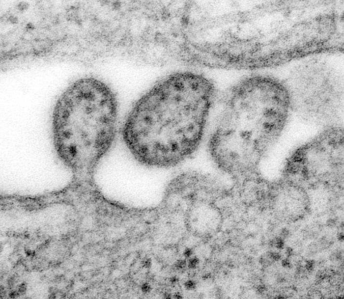
ID#: 8699 Description: This highly magnified transmission electron micrograph (TEM) depicted some of the ultrastructural details of a number of Lassa virus virions adjacent to some cell debris. The virus, a member of the virus family Arenaviridae, is a single-stranded RNA virus, and is zoonotic, or animal-borne that can be transmitted to humans. The illness, which occurs in West Africa, was discovered in 1969 when two missionary nurses died in Nigeria, West Africa. In areas of Africa where the disease is endemic (that is, constantly present), Lassa fever is a significant cause of morbidity and mortality. While Lassa fever is mild or has no observable symptoms in about 80% of people infected with the virus, the remaining 20% have a severe multisystem disease. Lassa fever is also associated with occasional epidemics, during which the case-fatality rate can reach 50%. Signs and symptoms of Lassa fever typically occur 1-3 weeks after the patient comes into contact with the virus. These include fever, retrosternal pain (pain behind the chest wall), sore throat, back pain, cough, abdominal pain, vomiting, diarrhea, conjunctivitis, facial swelling, proteinuria (protein in the urine), and mucosal bleeding. Neurological problems have also been described, including hearing loss, tremors, and encephalitis. Because the symptoms of Lassa fever are so varied and nonspecific, clinical diagnosis is often difficult. Approximately 15%-20% of patients hospitalized for Lassa fever die from the illness. However, overall only about 1% of infections with Lassa virus result in death. The death rates are particularly high for women in the third trimester of pregnancy, and for fetuses, about 95% of which die in the uterus of infected pregnant mothers. |
|||
| 来源 | http://phil.cdc.gov/PHIL_Images/8699/8699_lores.jpg | |||
| 作者 |
Content Providers(s): CDC/ C. S. Goldsmith, D. Auperin Photo Credit: C. S. Goldsmith Copyright Restrictions: None - This image is in the public domain and thus free of any copyright restrictions. As a matter of courtesy we request that the content provider be credited and notified in any public or private usage of this image. |
|||
| 授权 (二次使用本文件) |
|
文件历史
点击某个日期/时间查看对应时刻的文件。
| 日期/时间 | 缩略图 | 大小 | 用户 | 备注 | |
|---|---|---|---|---|---|
| 当前 | 2006年5月30日 (二) 16:56 | | 700 × 609(84 KB) | Patho(留言 | 贡献) | {{Information| |Description=ID#: 8699 Description: This highly magnified transmission electron micrograph (TEM) depicted some of the ultrastructural details of a number of Lassa virus virions adjacent to some cell debris. The virus, a member of the virus |
您不可以覆盖此文件。
文件用途
以下页面使用本文件:
全域文件用途
以下其他wiki使用此文件:
- ar.wikipedia.org上的用途
- arz.wikipedia.org上的用途
- bg.wikipedia.org上的用途
- ca.wikipedia.org上的用途
- cs.wikipedia.org上的用途
- da.wikipedia.org上的用途
- de.wikipedia.org上的用途
- de.wikibooks.org上的用途
- es.wikipedia.org上的用途
- fr.wikipedia.org上的用途
- he.wikipedia.org上的用途
- ja.wikipedia.org上的用途
- kk.wikipedia.org上的用途
- ko.wikipedia.org上的用途
- nl.wikipedia.org上的用途
- pt.wikipedia.org上的用途
- ru.wikipedia.org上的用途
- sl.wikipedia.org上的用途
- species.wikimedia.org上的用途
- uk.wikipedia.org上的用途
- www.wikidata.org上的用途
- zh.wikipedia.org上的用途