File:Late infantile metachromatic leukodystrophy cranial MRI.gif
Jump to navigation
Jump to search

Size of this preview: 339 × 598 pixels. Other resolutions: 136 × 240 pixels | 562 × 992 pixels.
Original file (562 × 992 pixels, file size: 437 KB, MIME type: image/gif)
Captions
Captions
Add a one-line explanation of what this file represents
| Description |
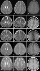
English: Cranial MRIs of our patients. a-c represents patient 1, d-f patient 2, g-i patient 3, j-l patient 4, and m-o patient 5. (a, d, g, j, m) Hypointense radially oriented stripes and dots seen within the hyperintense cerebral white matter (resembling tiger skin) on T2-weighted axial imaging. (b, e, h, k, n) Hypointense dots resembling leopard skin seen on T2-weighted axial imaging at the level of centrum ovale. (c, f, i, l, o) Iso to hyperintense dots seen in the cerebral white matter on T1-weighted imaging. This pattern of dysmyelination resembles the skin of tiger (radial stripes) and leopard (dots), the so-called tigroid and leopard pattern of dysmyelination in metachromatic leukodystrophy. |
| Date | |
| Source | Liaw, Hsiang-Ru, et al. "Late infantile metachromatic leukodystrophy: Clinical manifestations of five Taiwanese patients and Genetic features in Asia." Orphanet journal of rare diseases 10.1 (2015): 1. |
| Author | HR Liaw, HF Lee, CS Chi |
| Permission (Reusing this file) |
This article is distributed under the terms of the Creative Commons Attribution 4.0 International License |
This file is licensed under the Creative Commons Attribution 4.0 International license.
- You are free:
- to share – to copy, distribute and transmit the work
- to remix – to adapt the work
- Under the following conditions:
- attribution – You must give appropriate credit, provide a link to the license, and indicate if changes were made. You may do so in any reasonable manner, but not in any way that suggests the licensor endorses you or your use.
File history
Click on a date/time to view the file as it appeared at that time.
| Date/Time | Thumbnail | Dimensions | User | Comment | |
|---|---|---|---|---|---|
| current | 05:50, 21 July 2016 | | 562 × 992 (437 KB) | Filip em (talk | contribs) | {{Information |Description ={{en|1=Cranial MRIs of our patients. a-c represents patient 1, d-f patient 2, g-i patient 3, j-l patient 4, and m-o patient 5. (a, d, g, j, m) Hypointense radially oriented stripes and dots seen within the hyperintense ce... |
You cannot overwrite this file.
File usage on Commons
There are no pages that use this file.
File usage on other wikis
The following other wikis use this file:
- Usage on pl.wikipedia.org
- Usage on sr.wikipedia.org