File:Long bone assigned to Pterodactylus cuvieri.jpg
跳至導覽
跳至搜尋
預覽大小:800 × 241 像素。 其他解析度:320 × 96 像素 | 640 × 193 像素 | 1,024 × 308 像素 | 1,280 × 386 像素 | 4,740 × 1,428 像素。
原始檔案 (4,740 × 1,428 像素,檔案大小:4.46 MB,MIME 類型:image/jpeg)
說明
說明
添加單行說明來描述出檔案所代表的內容
摘要
[編輯]
| 描述 |
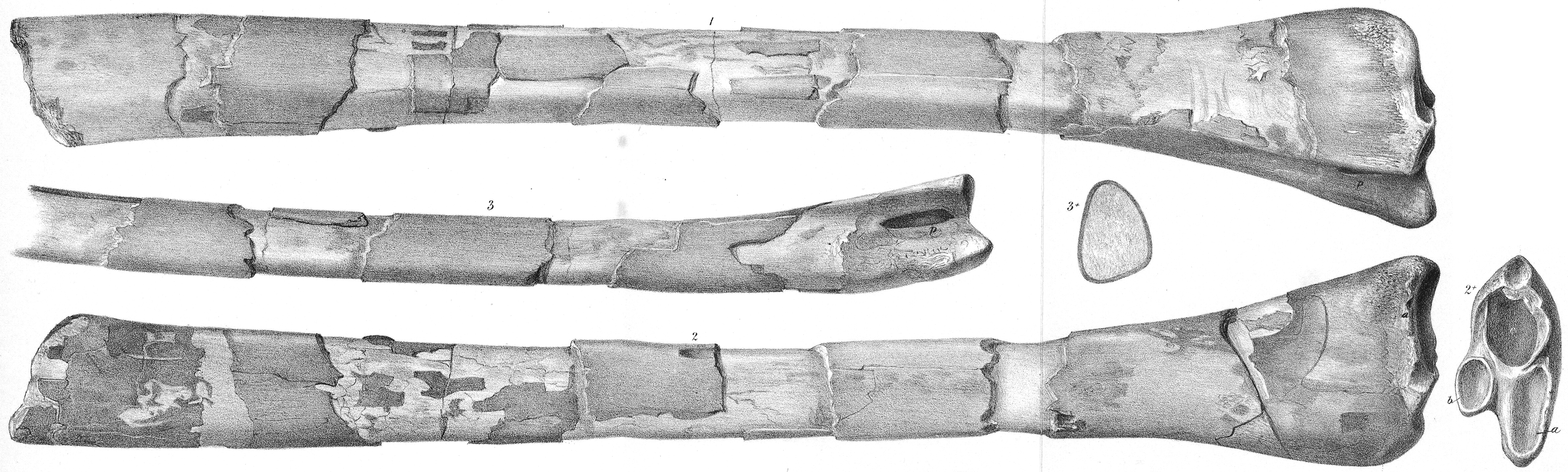
Figs. 1 and 2. Wing-phalanx bone of Pterodactylus Cuvieri. Fig. 2*. Articular end of ditto: a and b, articular surfaces; c, fractured surface leading to the cavity of the bone. Fig. 3. Portion of the narrowest side of the same bone, showing the pneumatic foramen at p. Fig. 3*. Section of the same bone four inches from the articular end, showing the thickness of its dense osseous wall, and the wide air-cavity. From the Burham Chalk-pit, Kent. In the Collection of J. Toulmin Smith, Esq. |
| 日期 | 在1849年到1884年之間 |
| 來源 | https://www.biodiversitylibrary.org/item/99821#page/25/mode/1up |
| 作者 | Joseph Dinkel |
授權條款
[編輯]
|
本作品在其來源國以及其他版權期限是作者逝世後70年或以下的國家與地區屬於公有領域。 | |
| 此作品無已知的著作權限制,亦不受所有相關和鄰接的權利限制。 | |
檔案歷史
點選日期/時間以檢視該時間的檔案版本。
| 日期/時間 | 縮圖 | 尺寸 | 用戶 | 備註 | |
|---|---|---|---|---|---|
| 目前 | 2021年2月16日 (二) 21:43 | 4,740 × 1,428(4.46 MB) | FunkMonk(對話 | 貢獻) | {{Information |Description=Figs. 1 and 2. Wing-bone of Pterodactylus Cuvieri. Fig. 2*. Articular end of ditto: a and b, articular surfaces; c, fractured surface leading to the cavity of the bone. Fig. 3. Portion of the narrowest side of the same bone, showing the pneumatic foramen at p. Fig. 3*. Section of the same bone four inches from the articular end, showing the thickness of its dense osseous wall, and the wide air-cavity. From the Burham Chalk-pit, Kent. In the Collection of J. Toulm... |
無法覆蓋此檔案。
檔案用途
沒有使用此檔案的頁面。
全域檔案使用狀況
以下其他 wiki 使用了這個檔案:
- en.wikipedia.org 的使用狀況
- fr.wikipedia.org 的使用狀況
- zh.wikipedia.org 的使用狀況