File:Macular corneal dystrophy hale colloidal iron stain.JPEG
Ir a la navegación
Ir a la búsqueda
No disponible a mayor resolución.
Macular_corneal_dystrophy_hale_colloidal_iron_stain.JPEG (500 × 500 píxeles; tamaño de archivo: 152 kB; tipo MIME: image/jpeg)
Leyendas
Leyendas
Añade una explicación corta acerca de lo que representa este archivo
| Descripción |
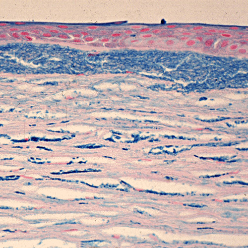
English: Macular corneal dystrophy. The abnormalities within the cornea are easily seen within the keratocytes and in a subepithelial extracellular location because they stain prominently with methods that demonstrate glycosaminoglycans. Hale colloidal iron stain. Klintworth Orphanet Journal of Rare Diseases 2009 4:7 doi:10.1186/1750-1172-4-7 Русский: Пятнистая дистрофия роговицы. Окрашивание гликозаминогликанов выявляет нарушения в кератоцитах стромы и во внеклеточном пространстве под эпителием роговицы. |
| Fecha | |
| Fuente | Corneal dystrophies |
| Autor | Klintworth GK. |
| Permiso (Reutilización de este archivo) |
© 2009 Klintworth; licensee BioMed Central Ltd. This is an Open Access article distributed under the terms of the Creative Commons Attribution License (https://creativecommons.org/licenses/by/2.0), which permits unrestricted use, distribution, and reproduction in any medium, provided the original work is properly cited. |
Este archivo está disponible bajo la licencia Creative Commons Atribución 2.0 Genérica.
- Eres libre:
- de compartir – de copiar, distribuir y transmitir el trabajo
- de remezclar – de adaptar el trabajo
- Bajo las siguientes condiciones:
- atribución – Debes otorgar el crédito correspondiente, proporcionar un enlace a la licencia e indicar si realizaste algún cambio. Puedes hacerlo de cualquier manera razonable pero no de manera que sugiera que el licenciante te respalda a ti o al uso que hagas del trabajo.
Historial del archivo
Haz clic sobre una fecha y hora para ver el archivo tal como apareció en ese momento.
| Fecha y hora | Miniatura | Dimensiones | Usuario | Comentario | |
|---|---|---|---|---|---|
| actual | 06:42 5 jul 2009 | | 500 × 500 (152 kB) | CopperKettle (discusión | contribs.) | {{Information |Description={{en|1=Macular corneal dystrophy. The abnormalities within the cornea are easily seen within the keratocytes and in a subepithelial extracellular location because they stain prominently with methods that demonstrate glycosaminog |
No puedes sobrescribir este archivo.
Usos del archivo
La siguiente página usa este archivo:
Uso global del archivo
Las wikis siguientes utilizan este archivo:
- Uso en de.wikipedia.org
- Uso en en.wikipedia.org
- Uso en es.wikipedia.org
- Uso en it.wikipedia.org
- Uso en ru.wikipedia.org
- Uso en tt.wikipedia.org
- Uso en www.wikidata.org