File:Mcn23702.jpg
From Wikimedia Commons, the free media repository
Jump to navigation
Jump to search

Size of this preview: 211 × 599 pixels. Other resolutions: 84 × 240 pixels | 169 × 480 pixels | 750 × 2,129 pixels.
Original file (750 × 2,129 pixels, file size: 986 KB, MIME type: image/jpeg)
File information
Structured data
Captions
Captions
Add a one-line explanation of what this file represents
Summary[edit]
| DescriptionMcn23702.jpg |
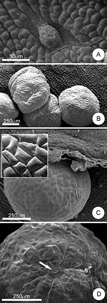
English: Scanning electron microscopy images of Cissus verticillata food bodies. (A) Locality where food bodies are formed on the stem; note the depression in which the FBs are formed in the midst of the papilose epidermal cells. (B) Food bodies in the nodal region of the stem. (C) Food body in the nodal region, partially protected by the stipule. Inset: detail of the epidermal cells of a food body; note the absence of any pores or ruptured areas of the cuticle. (D) Food body on a leaf, detail of the distal portion, demonstrating a stomata (arrow). |
| Date | |
| Source | https://www.ncbi.nlm.nih.gov/pmc/articles/PMC2707332/ |
| Author | Elder Antônio Sousa Paiva, Rafael Andrade Buono and Julio Antonio Lombardi |
Licensing[edit]
| Public domainPublic domainfalsefalse |
| This image is a work of the National Institutes of Health, part of the United States Department of Health and Human Services, taken or made as part of an employee's official duties. As a work of the U.S. federal government, the image is in the public domain.
|
 | |
| This file has been identified as being free of known restrictions under copyright law, including all related and neighboring rights. | ||
https://creativecommons.org/publicdomain/mark/1.0/PDMCreative Commons Public Domain Mark 1.0falsefalse
File history
Click on a date/time to view the file as it appeared at that time.
| Date/Time | Thumbnail | Dimensions | User | Comment | |
|---|---|---|---|---|---|
| current | 13:16, 4 March 2014 | 750 × 2,129 (986 KB) | Paul venter (talk | contribs) | {{Information |Description ={{en|1=Scanning electron microscopy images of Cissus verticillata food bodies. (A) Locality where food bodies are formed on the stem; note the depression in which the FBs are formed in the midst of the papilose epidermal... |
You cannot overwrite this file.
File usage on Commons
There are no pages that use this file.
File usage on other wikis
The following other wikis use this file:
- Usage on en.wikipedia.org
