File:Mitochondria, mammalian lung - TEM.jpg
Jump to navigation
Jump to search
No higher resolution available.
Mitochondria,_mammalian_lung_-_TEM.jpg (640 × 480 pixels, file size: 96 KB, MIME type: image/jpeg)
Captions
Captions
Add a one-line explanation of what this file represents
Summary[edit]
| Description |
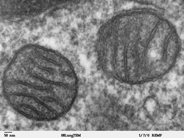
Transmission electron microscope image of a thin section cut through an area of mammalian lung tissue. The high magnification image shows two mitochondria. JEOL 100CX TEM |
| Source | |
| Author | Louisa Howard |
| Permission (Reusing this file) |
PD |
Licensing[edit]
| This work has been released into the public domain by its author, Louisa Howard. This applies worldwide. In some countries this may not be legally possible; if so: Louisa Howard grants anyone the right to use this work for any purpose, without any conditions, unless such conditions are required by law.
|
File history
Click on a date/time to view the file as it appeared at that time.
| Date/Time | Thumbnail | Dimensions | User | Comment | |
|---|---|---|---|---|---|
| current | 15:09, 16 May 2008 | | 640 × 480 (96 KB) | Vojtěch Dostál (talk | contribs) | Reverted to version as of 15:37, 5 October 2006, my fault |
| 12:51, 16 May 2008 |  | 640 × 433 (84 KB) | Vojtěch Dostál (talk | contribs) | {{Information |Description=Transmission electron microscope image of a thin section cut through an area of mammalian lung tissue. The high magnification image shows a mitochondria. JEOL 100CX TEM |Source= * http://remf.dartmouth.edu/imagesindex.html * h | |
| 12:47, 16 May 2008 |  | 640 × 453 (86 KB) | Vojtěch Dostál (talk | contribs) | {{Information |Description=Transmission electron microscope image of a thin section cut through an area of mammalian lung tissue. The high magnification image shows a mitochondria. JEOL 100CX TEM |Source= * http://remf.dartmouth.edu/imagesindex.html * h | |
| 15:37, 5 October 2006 |  | 640 × 480 (96 KB) | Patho (talk | contribs) | {{Information |Description=Transmission electron microscope image of a thin section cut through an area of mammalian lung tissue. The high magnification image shows a mitochondria. JEOL 100CX TEM |Source= * http://remf.dartmouth.edu/imagesindex.html * h |
You cannot overwrite this file.
File usage
The following 4 pages use this file:
.jpg)
Global file usage
The following other wikis use this file:
- Usage on ar.wikipedia.org
- Usage on az.wiktionary.org
- Usage on be.wikipedia.org
- Usage on bg.wikipedia.org
- Usage on bn.wikipedia.org
- Usage on br.wikipedia.org
- Usage on bs.wikipedia.org
- Usage on ca.wikipedia.org
- Usage on cdo.wikipedia.org
- Usage on da.wikipedia.org
- Usage on de.wikibooks.org
- Usage on el.wikipedia.org
- Usage on en.wikipedia.org
- Usage on en.wikibooks.org
- Usage on en.wikiversity.org
- User:Jtwsaddress42/Projects/Project 1
- User:Jtwsaddress42/Projects/Project 1/Parts
- User:Jtwsaddress42/Projects/Project 1/Parts/Part 3
- User:Jtwsaddress42/Projects/Project 1/Chapters/Chapter 10
- User:Jtwsaddress42/Projects/Project 1/Sections/Chapter 10/Phase II - The Oxygen Crisis and the Rise of the Aerobic Bioshphere (1.9-0.95 bya)
- User:Jtwsaddress42/Clade
- User:Jtwsaddress42/Clade/Gracilicutes to Proteobacteria
- Usage on en.wiktionary.org
- Usage on es.wikipedia.org
- Usage on et.wikipedia.org
- Usage on eu.wikipedia.org
- Usage on ext.wikipedia.org
- Usage on fa.wikipedia.org
View more global usage of this file.