File:Mitosis cells sequence.svg
Jump to navigation
Jump to search

Size of this PNG preview of this SVG file: 774 × 115 pixels. Other resolutions: 320 × 48 pixels | 640 × 95 pixels | 1,024 × 152 pixels | 1,280 × 190 pixels | 2,560 × 380 pixels.
Original file (SVG file, nominally 774 × 115 pixels, file size: 459 KB)
Captions
Captions
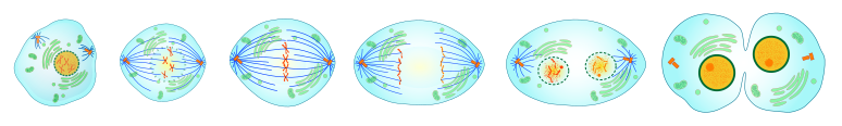
The phases of mitosis, Interphase, prophase, metaphase, anaphase, telophase, plus cytokinesis
Summary[edit]
| Description | A series of cell diagrams showing the mitosis division of eukaryotic cells. | ||
| Date | |||
| Source |
Own work. The cells are extracted from: |
||
| Author | LadyofHats | ||
| Permission (Reusing this file) |
|
||
| Other versions |
Derivative works of this file: |


File history
Click on a date/time to view the file as it appeared at that time.
| Date/Time | Thumbnail | Dimensions | User | Comment | |
|---|---|---|---|---|---|
| current | 10:00, 10 September 2008 | 774 × 115 (459 KB) | LadyofHats (talk | contribs) | {{Information |Description= a serie of cells showing the mitosis divition of eucaryotic cells |Source=own work, the cells are extracted from: <gallery> Image:Prophase procariotic mitosis.svg Image:Prometaphase procariotic mitosis.svg Image:Metaphase proca |
You cannot overwrite this file.
File usage
The following 3 pages use this file:
Global file usage
The following other wikis use this file:
- Usage on ast.wikipedia.org
- Usage on cy.wikipedia.org
- Usage on en.wikipedia.org
- Usage on es.wikipedia.org
- Usage on eu.wikipedia.org
- Usage on fr.wiktionary.org
- Usage on nl.wikibooks.org
- Usage on sq.wikipedia.org