File:Monocot Root Stele in Lilium (36045154705).jpg

元のファイル (3,264 × 1,840 ピクセル、ファイルサイズ: 1.07メガバイト、MIME タイプ: image/jpeg)
キャプション
キャプション
概要
[編集].jpg&action=edit§ion=1)
| 解説 |
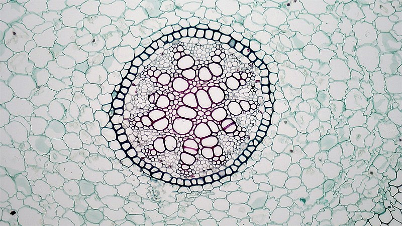
cross section:Lilium root common name: lily magnification: 100x <a href="http://blogs.berkshirecc.edu/bccoer" rel="nofollow">Berkshire Community College Bioscience Image Library</a> The single layered epidermis lacks a cuticle. Root hairs appear as unicellular extensions of epidermal cells. The cortex is well-developed and divided into two zones; a narrow outer layer of closely packed smaller parenchyma cells and a wide inner layer of open larger aerenchyma cells The inmost area of the cortex is bounded by a prominent endodermis of cells whose walls are heavily suberized, marking the Casparian stip. The outer stele is bound is a pericycle of one layer thin walled parenchyma cells. The vascular tissues consist of radiating arms of xylem and phloem with phloem forming strands near the periphery of the vascular cylinder. The xylem is exarch: meaning the earlier smaller protoxylem is found towards the periphery and younger larger metaxylem to the center of the stem. Phloem tissues of sieve tubes, companion cells and phloem parenchyma are also exarch. The older protophloem is found towards the periphery and metaphloem towards the center of the stem. Phloem parenchyma may become sclerenchymized in older roots. Vascular cambium is absent, preventing secondary growth of the root. The central of the stele is occupied by pith containing thin walled starch containing parenchyma cells that may become sclerenchymized in older roots. |
| 日付 | |
| 原典 | Monocot Root: Stele in Lilium |
| 作者 | Berkshire Community College Bioscience Image Library |
ライセンス
[編集].jpg&action=edit§ion=2)
| このファイルはクリエイティブ・コモンズ CC0 1.0 全世界 パブリック・ドメイン提供のもとで利用可能にされています。 | |
| ある作品に本コモンズ証を関連づけた者は、その作品について世界全地域において著作権法上認められる、その者が持つすべての権利(その作品に関する権利や隣接する権利を含む。)を、法令上認められる最大限の範囲で放棄して、パブリック・ドメインに提供しています。
この作品は、たとえ営利目的であっても、許可を得ずに複製、改変・翻案、配布、上演・演奏することが出来ます。 |
| この画像は当初、bccoer によって Flickr の https://flickr.com/photos/146824358@N03/36045154705 に投稿されたものです。2019-10-05、FlickreviewR 2 ボットによってレビューされ、cc-zero のライセンスで提供されていることが確認されました。 |
Files from Berkshire Community College Bioscience Image Library Flickr stream uploaded by Netha Hussain
ファイルの履歴
過去の版のファイルを表示するには、その版の日時をクリックしてください。
| 日付と時刻 | サムネイル | 寸法 | 利用者 | コメント | |
|---|---|---|---|---|---|
| 現在の版 | 2019年10月5日 (土) 19:40 | | 3,264 × 1,840 (1.07メガバイト) | Netha Hussain (トーク | 投稿記録) | Transferred from Flickr via #flickr2commons |
このファイルは上書きできません。
ファイルの使用状況
このファイルを使用しているページはありません。
.jpg&oldid=497430399)