File:MultiPhotonExcitation-Fig10-doi10.1186slash1475-925X-5-36-clipping.JPEG
Salta a la navegació
Salta a la cerca
No hi ha cap versió amb una resolució més gran.
MultiPhotonExcitation-Fig10-doi10.1186slash1475-925X-5-36-clipping.JPEG (714 × 467 píxels, mida del fitxer: 81 Ko, tipus MIME: image/jpeg)
Llegendes
Llegendes
Afegeix una explicació d'una línia del que representa aquest fitxer
Resum[modifica]
| Descripció |
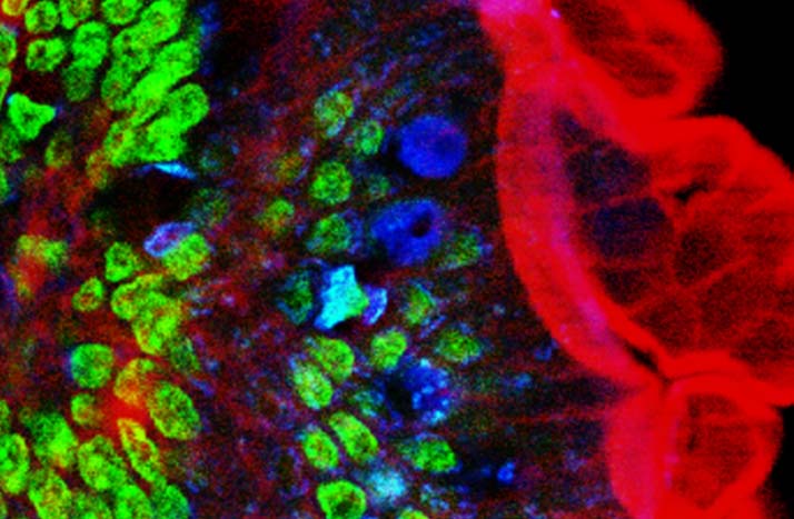
English: Original figure legend: Multiple fluorescence 2PE imaging. 2PE multiple fluorescence image from a 16 μm cryostat section of mouse intestine stained with a combination of fluorescent stains (F-24631, Molecular Probes). Alexa Fluor 350 wheat germ agglutinin, a blue-fluorescent lectin, was used to stain the mucus of goblet cells. The filamentous actin prevalent in the brush border was stained with red-fluorescent Alexa Flu or 568 phalloidin. Finally, the nuclei were stained with SYTOX ® Green nucleic acid stain. Imaging has been performed at 780 nm, 100 x 1.4 NA Leica objective, using a Chameleon XR ultrafast Ti-Sapphire laser (Coherent Inc., USA) coupled at LAMBS-MicroScoBio with a Spectral Confocal Laser Scanning Microscope, Leica SP2-AOBS.
Deutsch: Zweiphotonenaufnahme an einem Schnitt durch einen Mausdarm. Zellkerne in grün, Schleim der Becherzellen in blau, Aktin (Phalloidin-Färbung) in rot. Anregung erfolgte bei 780 nm durch einen Titan:Saphir-Laser.
Français : légende originale de l'image : imagerie en fluorescenc emultiple 2PE d'une section de 16 µm de cryostat d'intestin de souris coloré avec une combinaison de colorants fluorescents (F-24631, Molecular Probes). l'Alexa Fluor 350 d'agglutinine degerme de blé, une lectine bleu fluorescente, a été utilisée pour colorer le mucus des cellules caliciformes. L'actine filamenteuse a été colorée avec du rouge fluorescent (Alexa Flu ou phalloïdine 568). Enfin, les noyaux ont été colorés avec un autre colorant (SYTOX ® Green nucleic acid stain). L'image a été faite à 780 nm, avec un objectif Leica 100 x 1,4 NA, en utilisant un éclairage laser (Chameleon XR ultrafast Ti-Sapphire laser (Coherent Inc., USA) ) couplé à un microscope LAMBS-MicroScoBio (Spectral Confocal Laser Scanning Microscope, Leica SP2-AOBS). |
| Data | Original version: 6 June 2006. Clipping: 4. March 2009. |
| Font |
Multi-photon excitation microscopy. BioMedical Engineering OnLine, 2006, 5:36. |
| Autor |
Alberto Diaspro, Paolo Bianchini, Giuseppe Vicidomini, Mario Faretta, Paola Ramoino and Cesare Usai. |
| Permís (Com reutilitzar aquest fitxer) |
Aquest fitxer està disponible sota la llicència Creative Commons Reconeixement 2.0 Genèrica.
|
| Altres versions | For unclipped version see below |
All images uploaded from this article about multi-photon and two-photon-microscopy:


Historial del fitxer
Cliqueu una data/hora per veure el fitxer tal com era aleshores.
| Data/hora | Miniatura | Dimensions | Usuari/a | Comentari | |
|---|---|---|---|---|---|
| actual | 20:57, 4 març 2009 | | 714 × 467 (81 Ko) | Dietzel65 (discussió | contribucions) | == Beschreibung == {{Information |Description={{en|1=Original figure legend: ''Multiple fluorescence 2PE imaging. 2PE multiple fluorescence image from a 16 μm cryostat section of mouse intestine stained with a combination of fluorescent stains (F-24631, |
No podeu sobreescriure aquest fitxer.
Ús del fitxer
Les 2 pàgines següents utilitzen aquest fitxer:
Ús global del fitxer
Utilització d'aquest fitxer en altres wikis:
- Utilització a ar.wikipedia.org
- Utilització a ca.wikipedia.org
- Utilització a de.wikipedia.org
- Utilització a en.wikipedia.org
- Utilització a es.wikipedia.org
- Utilització a fr.wikipedia.org
- Utilització a it.wikipedia.org
- Utilització a outreach.wikimedia.org
- Utilització a uk.wikipedia.org
- Utilització a zh.wikipedia.org