File:MultiPhotonExcitation-Fig10-doi10.1186slash1475-925X-5-36-clipping.JPEG
出自Wikimedia Commons
跳至導覽
跳至搜尋
MultiPhotonExcitation-Fig10-doi10.1186slash1475-925X-5-36-clipping.JPEG (714 × 467 像素,檔案大小:81 KB,MIME 類型:image/jpeg)
檔案資訊
結構化資料
說明
說明
添加單行說明來描述出檔案所代表的內容
摘要
[編輯]| 描述MultiPhotonExcitation-Fig10-doi10.1186slash1475-925X-5-36-clipping.JPEG |
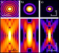
English: Original figure legend: Multiple fluorescence 2PE imaging. 2PE multiple fluorescence image from a 16 μm cryostat section of mouse intestine stained with a combination of fluorescent stains (F-24631, Molecular Probes). Alexa Fluor 350 wheat germ agglutinin, a blue-fluorescent lectin, was used to stain the mucus of goblet cells. The filamentous actin prevalent in the brush border was stained with red-fluorescent Alexa Flu or 568 phalloidin. Finally, the nuclei were stained with SYTOX ® Green nucleic acid stain. Imaging has been performed at 780 nm, 100 x 1.4 NA Leica objective, using a Chameleon XR ultrafast Ti-Sapphire laser (Coherent Inc., USA) coupled at LAMBS-MicroScoBio with a Spectral Confocal Laser Scanning Microscope, Leica SP2-AOBS.
Deutsch: Zweiphotonenaufnahme an einem Schnitt durch einen Mausdarm. Zellkerne in grün, Schleim der Becherzellen in blau, Aktin (Phalloidin-Färbung) in rot. Anregung erfolgte bei 780 nm durch einen Titan:Saphir-Laser.
Français : légende originale de l'image : imagerie en fluorescenc emultiple 2PE d'une section de 16 µm de cryostat d'intestin de souris coloré avec une combinaison de colorants fluorescents (F-24631, Molecular Probes). l'Alexa Fluor 350 d'agglutinine degerme de blé, une lectine bleu fluorescente, a été utilisée pour colorer le mucus des cellules caliciformes. L'actine filamenteuse a été colorée avec du rouge fluorescent (Alexa Flu ou phalloïdine 568). Enfin, les noyaux ont été colorés avec un autre colorant (SYTOX ® Green nucleic acid stain). L'image a été faite à 780 nm, avec un objectif Leica 100 x 1,4 NA, en utilisant un éclairage laser (Chameleon XR ultrafast Ti-Sapphire laser (Coherent Inc., USA) ) couplé à un microscope LAMBS-MicroScoBio (Spectral Confocal Laser Scanning Microscope, Leica SP2-AOBS). |
| 日期 | Original version: 6 June 2006. Clipping: 4. March 2009. |
| 來源 |
Multi-photon excitation microscopy. BioMedical Engineering OnLine, 2006, 5:36. |
| 作者 |
Alberto Diaspro, Paolo Bianchini, Giuseppe Vicidomini, Mario Faretta, Paola Ramoino and Cesare Usai. |
| 授權許可 (重用此檔案) |
此檔案採用創用CC 姓名標示 2.0 通用版授權條款。
|
| 其他版本 | For unclipped version see below |
All images uploaded from this article about multi-photon and two-photon-microscopy:
檔案歷史
點選日期/時間以檢視該時間的檔案版本。
| 日期/時間 | 縮圖 | 尺寸 | 用戶 | 備註 | |
|---|---|---|---|---|---|
| 目前 | 2009年3月4日 (三) 20:57 |  | 714 × 467(81 KB) | Dietzel65(對話 | 貢獻) | == Beschreibung == {{Information |Description={{en|1=Original figure legend: ''Multiple fluorescence 2PE imaging. 2PE multiple fluorescence image from a 16 μm cryostat section of mouse intestine stained with a combination of fluorescent stains (F-24631, |
無法覆蓋此檔案。
檔案用途
下列2個頁面有用到此檔案:
全域檔案使用狀況
以下其他 wiki 使用了這個檔案:
- ar.wikipedia.org 的使用狀況
- ca.wikipedia.org 的使用狀況
- de.wikipedia.org 的使用狀況
- en.wikipedia.org 的使用狀況
- es.wikipedia.org 的使用狀況
- fr.wikipedia.org 的使用狀況
- it.wikipedia.org 的使用狀況
- outreach.wikimedia.org 的使用狀況
- uk.wikipedia.org 的使用狀況
- zh.wikipedia.org 的使用狀況
詮釋資料
此檔案中包含其他資訊,這些資訊可能是由數位相機或掃描器在建立或數位化過程中所新增的。若檔案自原始狀態已被修改,一些詳細資料可能無法完整反映出已修改的檔案。
| 作者 | TCS User |
|---|---|
| 方位 | 標準 |
| 水平解析度 | 72 dpi |
| 垂直解析度 | 72 dpi |
| 使用軟體 | Adobe Photoshop 7.0 |
| 檔案修改日期時間 | 2009年3月4日 (三) 21:47 |
| 亮度與彩度位置 | 中間 |
| Exif 版本 | 2.2 |
| 色彩空間 | 顏色未校準 |


