File:Myocardial bridges.jpg
Jump to navigation
Jump to search
No higher resolution available.
Myocardial_bridges.jpg (631 × 537 pixels, file size: 55 KB, MIME type: image/jpeg)
Captions
Captions
Add a one-line explanation of what this file represents
| Description |
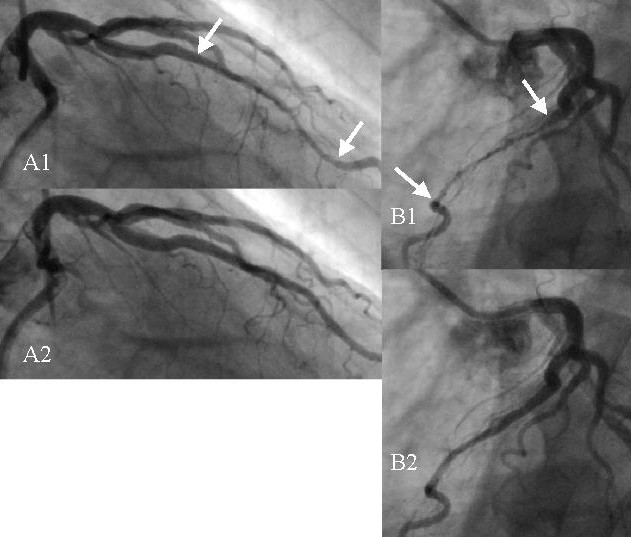
English: Angiographic images showing a bridge on the left anterior descending coronary artery (LAD) in a male patient of 65 years. A1) Right anterior oblique view taken at end systole. The compressed vessel segment is indicated by the two arrows. B1) Left anterior oblique view taken nearly at the same instant. A2) Same view as in A1, but taken 133 ms later. The tunneled segment is no longer compressed. B2) Same view as in B1 but 133 ms later. |
| Date | |
| Source | Doriot et al. Theoretical Biology and Medical Modelling 2007 4:29 doi:10.1186/1742-4682-4-29 |
| Author | Doriot |
This file is licensed under the Creative Commons Attribution 2.0 Generic license.
- You are free:
- to share – to copy, distribute and transmit the work
- to remix – to adapt the work
- Under the following conditions:
- attribution – You must give appropriate credit, provide a link to the license, and indicate if changes were made. You may do so in any reasonable manner, but not in any way that suggests the licensor endorses you or your use.
File history
Click on a date/time to view the file as it appeared at that time.
| Date/Time | Thumbnail | Dimensions | User | Comment | |
|---|---|---|---|---|---|
| current | 02:34, 29 April 2008 | | 631 × 537 (55 KB) | Filip em (talk | contribs) | {{Information |Description=Angiographic images showing a bridge on the left anterior descending coronary artery (LAD) in a male patient of 65 years. A1) Right anterior oblique view taken at end systole. The compressed vessel segment is indicated by the tw |
You cannot overwrite this file.
File usage on Commons
There are no pages that use this file.
File usage on other wikis
The following other wikis use this file:
- Usage on ar.wikipedia.org
- Usage on en.wikipedia.org
- Usage on fr.wikipedia.org
- Usage on he.wikipedia.org
- Usage on pl.wikipedia.org