File:Nidovirales life cycle.webp
Jump to navigation
Jump to search

Size of this PNG preview of this WEBP file: 800 × 591 pixels. Other resolutions: 320 × 236 pixels | 640 × 472 pixels | 1,024 × 756 pixels | 1,280 × 945 pixels | 2,560 × 1,890 pixels | 3,747 × 2,766 pixels.
Original file (3,747 × 2,766 pixels, file size: 733 KB, MIME type: image/webp)
Captions
Captions
Life cycle of positive sense RNA virus (Nidovirales)
Summary
[edit]
| Description |
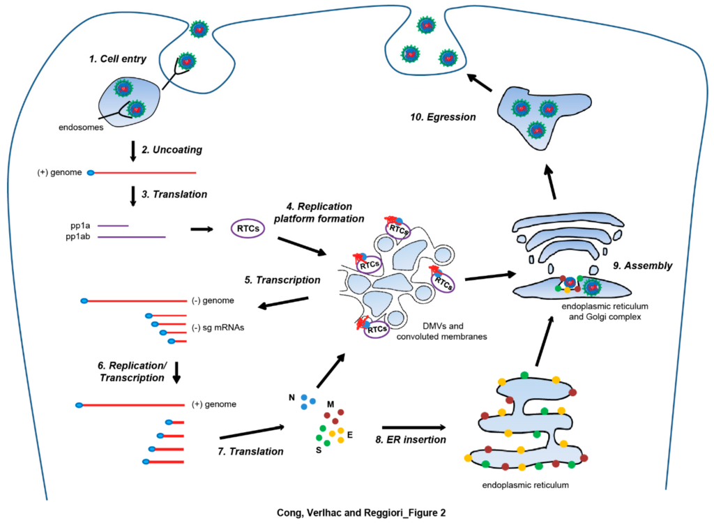
English: Generalization of Nidovirales life cycle, based on the information acquired studying Arteriviruses and Coronaviruses. Infection starts with the binding of the viral particle to a cell surface receptor and subsequent cell entry through membrane fusion in endosomes upon endocytosis (step 1). Translation of the released genomic RNA (gRNA) yields replicase polyproteins (step 2), i.e., polyprotein 1a (pp1a) and polyprotein 1ab (pp1ab), which undergo autoproteolytic processing to generate nonstructural proteins that assemble into replication-transcription complexes (RTCs). The RTCs are part of a complex membranous network composed of double membrane vesicles (DMVs) and convoluted membranes (step 4). The RTCs first engage in minus-strand RNA synthesis to produce both single strand full-length and subgenomic (sg) minus-strand RNAs (step 5). Subsequently, they use sg mRNAs as templates for the production of the gRNA and plus-strand sg mRNAs required to express the structural protein genes (step 6). Newly synthesized S, E, and M structural proteins are inserted in the endoplasmic reticulum (ER) (steps 7 and 8), whereas the N nucleocapsides are translated and oligomerize in the cytosol, where they interact with RTCs and associate with the gRNA to form the ribonucleoprotein complexes (step 7). Virion assembly takes place in the ER and/or Golgi (step 9), and involves the inward budding of the limiting membrane of these compartments, which is triggered by the interaction between the structural proteins and the ribonucleoprotein complexes. Mature virions are released extracellularly by exocytosis (step 10). |
| Date | |
| Source | https://www.mdpi.com/1999-4915/9/7/182/htm |
| Author | Yingying Cong, Pauline Verlhac, and Fulvio Reggiori |
Licensing
[edit]
This file is licensed under the Creative Commons Attribution 4.0 International license.
- You are free:
- to share – to copy, distribute and transmit the work
- to remix – to adapt the work
- Under the following conditions:
- attribution – You must give appropriate credit, provide a link to the license, and indicate if changes were made. You may do so in any reasonable manner, but not in any way that suggests the licensor endorses you or your use.
File history
Click on a date/time to view the file as it appeared at that time.
| Date/Time | Thumbnail | Dimensions | User | Comment | |
|---|---|---|---|---|---|
| current | 08:52, 9 April 2020 | | 3,747 × 2,766 (733 KB) | Guest2625 (talk | contribs) | Uploaded a work by Yingying Cong, Pauline Verlhac, and Fulvio Reggiori from https://www.mdpi.com/1999-4915/9/7/182/htm with UploadWizard |
You cannot overwrite this file.
File usage on Commons
There are no pages that use this file.
File usage on other wikis
The following other wikis use this file:
- Usage on en.wikipedia.org
- Usage on ja.wikipedia.org