File:PBP catalysis.svg

پروندهٔ اصلی (پروندهٔ اسویجی، با ابعاد ۱٬۱۴۲ × ۱٬۵۶۷ پیکسل، اندازهٔ پرونده: ۱٫۴۴ مگابایت)
گزینهها
عنوان
خلاصه
[ویرایش]| توضیح |
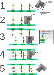
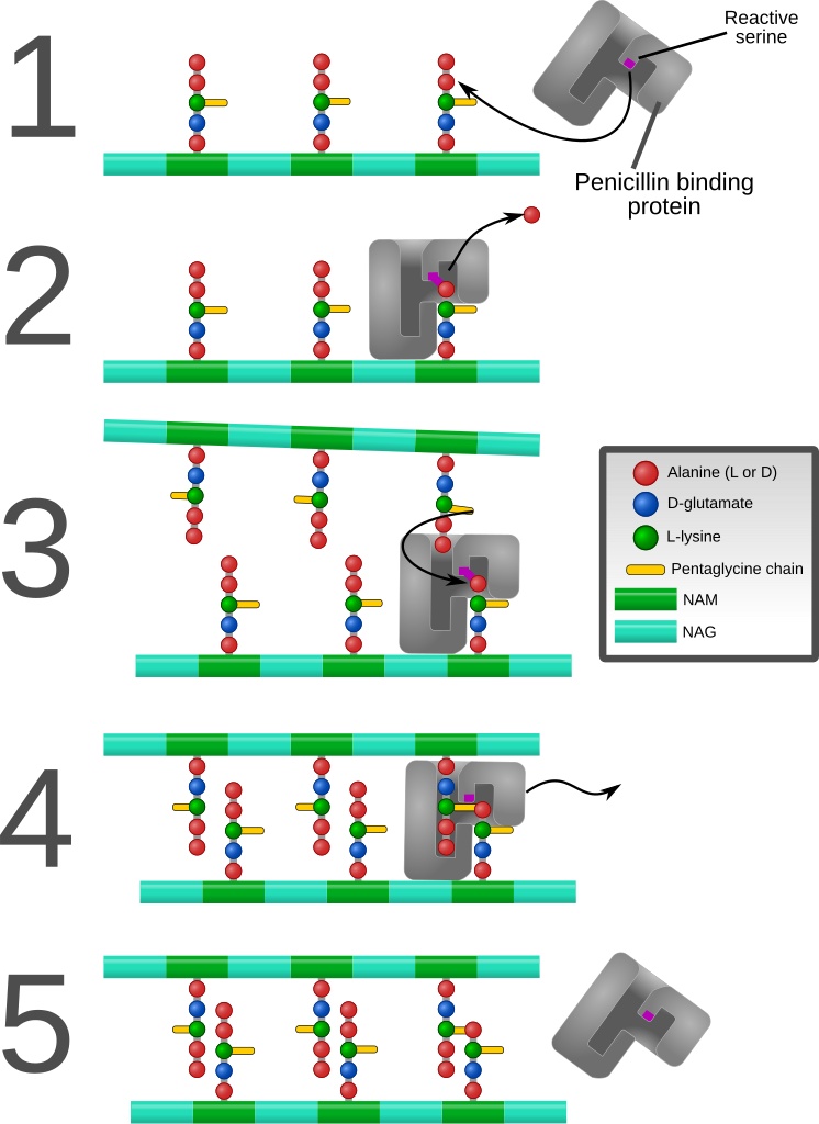
English: Diagram depicting formation of cross-links in the bacterial cell wall by a penicillin binding protein (PBP, an enzyme).
1. The bacterial cell wall consists of strands of repeating N-acetylglucosamine (NAG) and N-acetylmuramic acid (NAM) subunits. The NAM subunits have short peptide chains attached to them. (The exact composition of these can vary. The proximal alanine is usually L-ala and the distal two are usually D-ala.) These chains, in turn, are bound to chains of 5 glycine residues that will be used in cross-linking. 2. The PBP forms a bond with the peptide side chain at the second most distal alanine residue. This displaces the most distal alanine residue. 3. Another strand of bacterial cell wall arrives. The free end of one of the pentaglycine chains displaces the PBP and forms a bond with the terminal alanine on the other strand. 4. After being displaced, the PBP diffuses away. 5. The formation of one cross-link is complete. |
| تاریخ | |
| منبع | اثر شخصی |
| پدیدآور | Mcstrother |
| دیگر نسخهها |
[]
|


اجازهنامه
[ویرایش]
- شما اجازه دارید:
- برای به اشتراک گذاشتن – برای کپی، توزیع و انتقال اثر
- تلفیق کردن – برای انطباق اثر
- تحت شرایط زیر:
- انتساب – شما باید اعتبار مربوطه را به دست آورید، پیوندی به مجوز ارائه دهید و نشان دهید که آیا تغییرات ایجاد شدهاند یا خیر. شما ممکن است این کار را به هر روش منطقی انجام دهید، اما نه به هر شیوهای که پیشنهاد میکند که مجوزدهنده از شما یا استفادهتان حمایت کند.
تاریخچهٔ پرونده
روی تاریخ/زمانها کلیک کنید تا نسخهٔ مربوط به آن هنگام را ببینید.
| تاریخ/زمان | بندانگشتی | ابعاد | کاربر | توضیح | |
|---|---|---|---|---|---|
| کنونی | ۹ سپتامبر ۲۰۱۱، ساعت ۱۹:۲۶ | | ۱٬۱۴۲ در ۱٬۵۶۷ (۱٫۴۴ مگابایت) | Mcstrother (بحث | مشارکتها) | Major revision. Corrected inaccuracies in previous image. |
| ۳ مهٔ ۲۰۱۱، ساعت ۰۴:۱۵ |  | ۱٬۱۳۹ در ۱٬۰۶۲ (۸۵۰ کیلوبایت) | Mcstrother (بحث | مشارکتها) | Changed all fonts to Liberation Sans | |
| ۱۰ آوریل ۲۰۱۱، ساعت ۰۳:۴۶ |  | ۱٬۱۳۹ در ۱٬۰۶۲ (۸۵۰ کیلوبایت) | Mcstrother (بحث | مشارکتها) | Changed color of carbohydrate chain. | |
| ۷ مارس ۲۰۱۱، ساعت ۰۳:۳۰ |  | ۱٬۱۳۹ در ۱٬۰۶۲ (۸۳۵ کیلوبایت) | Mcstrother (بحث | مشارکتها) | {{Information |Description ={{en|1=Diagram depicting formation of cross-links in the bacterial cell wall by a penicillin binding protein (PBP, an enzyme). 1. The bacterial cell wall consists of strands of repeating N-acetylglucosamine (NAG) and N-ace |
نمیتوانید این پرونده را رونویسی کنید.
کاربرد پرونده
صفحههای زیر از این تصویر استفاده میکنند:
کاربرد سراسری پرونده
ویکیهای دیگر زیر از این پرونده استفاده میکنند:
- کاربرد در en.wikipedia.org
- کاربرد در es.wikipedia.org
- کاربرد در fa.wikipedia.org
- کاربرد در ga.wikipedia.org
- کاربرد در gl.wikipedia.org
- کاربرد در hu.wikipedia.org
- کاربرد در it.wikipedia.org
- کاربرد در mk.wikipedia.org
- کاربرد در th.wikipedia.org