File:PZSL1889Plate46.png
Original file (1,903 × 3,026 pixels, file size: 5.5 MB, MIME type: image/png)
Captions
Captions
Summary
[edit]
| Description |
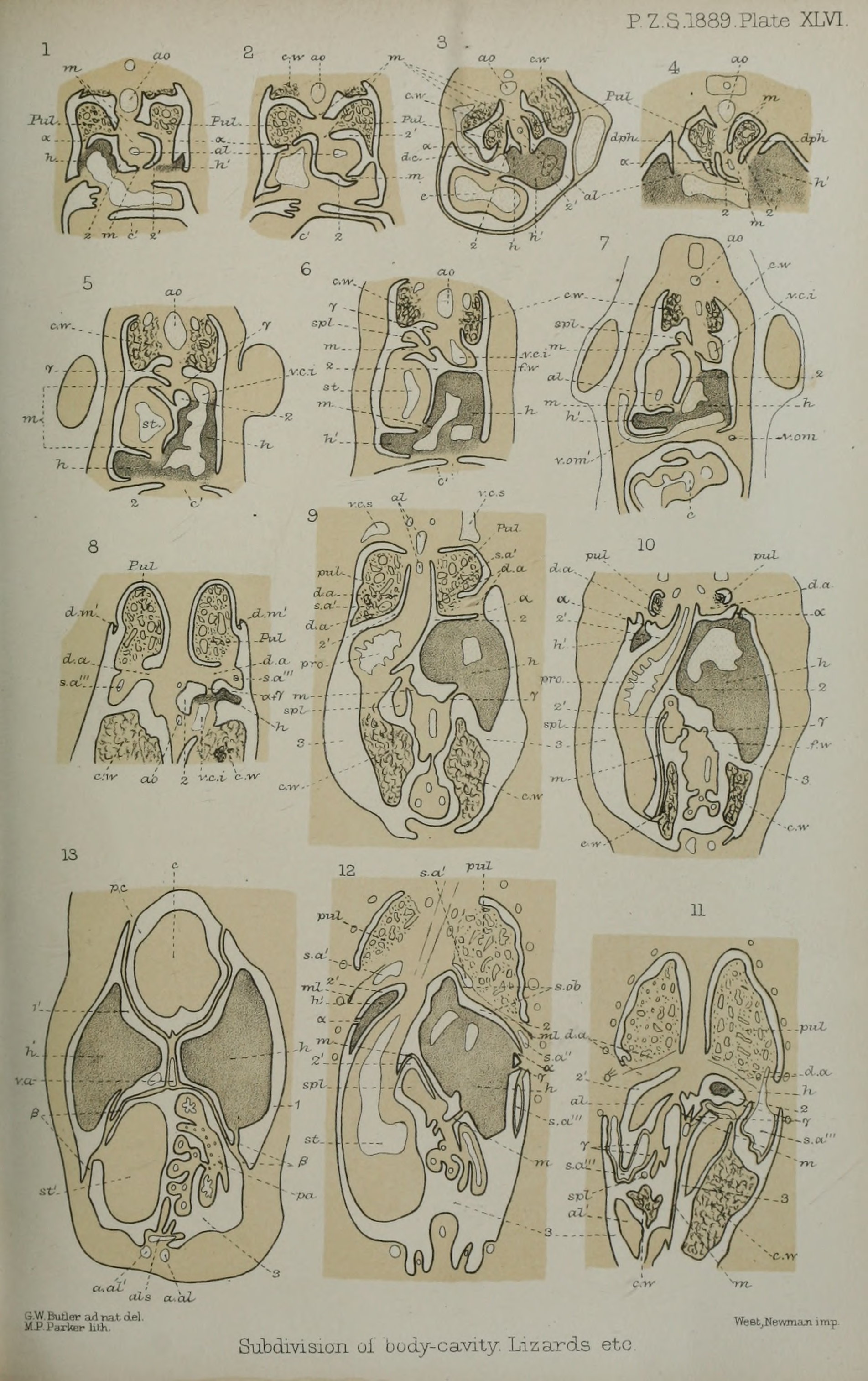
1-2, 5-13: Gallus gallus domesticus = Gallus gallus domesticus (Linnaeus, 1758) English: Microtome sections of vertebrate embryos:
a.al = right allantoic artery, a.al' = left allantoic artery, al = alimentary canal, als = allantois, ao = aorta dorsalis, c = heart, c' = heart wall, c.w = Wolffian body, d.a = diaphragm (avian), d.c = common cardinal vein (duct of Cuvier), d.m' = ligament of Müllerian duct, dph = diaphragm (mammalian), f.w = omental foramen (foramen of Winslow), h = right liver lobe, h' = left liver lobe, m = median thoracic septum and abdominal mesentery and ligaments around alimentary canal, ml = diaphragm muscle, pa = pancreas, pc = pericardium, pro = proventriculus, pul/Pul = lung, s.a' = anterior diaphragmatic air sac, s.a'' = posterior diaphragmatic air sac, s.a''' = abdominal air sac, s.ob = oblique septum, spl = spleen, [st. = stomach cavity, st.' = stomach wall,] v.a = anterior abdominal vein (left allantoic vein), v.c.i = vena cava inferior, v.c.s = vena cava superior, v.om = right omphalo-mesenteric vein, v.om' = left omphalo-mesenteric vein, 1 = right ventral liver sac, 1' = left ventral liver sac, 2 = right pulmohepatic recess, 2' = left pulmohepatic recess, 3 = peritoneal cavity, α = pulmohepatic ligament, β = omental septum (ventral portion of posthepatic septum), γ = oblique abdominal septum (antero-dorsal portion of posthepatic septum). |
||||
| Date | (published 1890) | ||||
| Source | Proceedings of the Zoological Society of London (vol. 1889 issue 4, plate XLVI) | ||||
| Author | G.W.Butler & M.P.Parker | ||||
| Permission (Reusing this file) |
|
||||
File history
Click on a date/time to view the file as it appeared at that time.
| Date/Time | Thumbnail | Dimensions | User | Comment | |
|---|---|---|---|---|---|
| current | 04:35, 7 June 2012 | | 1,903 × 3,026 (5.5 MB) | Dysmorodrepanis (talk | contribs) |
You cannot overwrite this file.
File usage on Commons
The following page uses this file: