File:Paracentrotus lividus ciliogenesis.jpg
Jump to navigation
Jump to search

Size of this preview: 424 × 599 pixels. Other resolutions: 170 × 240 pixels | 340 × 480 pixels | 891 × 1,259 pixels.
Original file (891 × 1,259 pixels, file size: 1.43 MB, MIME type: image/jpeg)
Captions
Captions
Add a one-line explanation of what this file represents
Summary[edit]
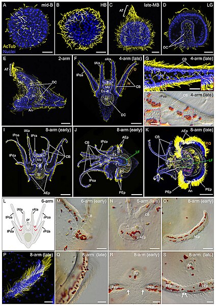
| Description | FIGURE 8. Ciliogenesis in Paracentrotus lividus during the embryonic and larval periods. Developmental stages are as follows: (A) mid-blastula stage (mid-B); (B) hatched blastula stage (HB); (C) late mesenchyme blastula stage (late-MB); (D) late gastrula stage (LG); (E) 2-arm pluteus stage (2-arm); (F–H) 4-arm pluteus stage (4-arm); (I–K, O–S) 8-arm pluteus stage (8-arm); (L–N) 6-arm pluteus stage (6-arm). The use of (early) or (late) associated with the stage names simply highlights here more specific periods during the 4-, 6- or 8-arm pluteus stages. In (A–G,I–K,P), images are maximum intensity projections of confocal z-stacks of embryos and larvae co-labeled for acetylated α-tubulin (cilia; yellow) and DNA (nuclei; blue), and they correspond to projections of the entire specimen, except for (D) that is a cross-section through the embryo. In (H,M–O,Q–S), images were acquired using light microscopy. (L) Schematics of a larva at the 6-arm pluteus stage illustrating in red the areas of the ciliary band that will bud to form the epaulettes. In (A–D), embryos are in lateral view with the animal pole up. In (E,J,K), larvae are in left view, with the anterior side up and the ventral side left. In (F,I), larvae are in anterior view, with the ventral side up. (G,H) Close-ups of the ciliary band of a late 4-arm pluteus stage larva, corresponding to the regions outlined by orange boxes in (F). ((G) inset, (H) inset), Close-ups of the ciliary band to highlight the distribution of cuboidal cells and their associated cilia. (M–O) Close-ups of a ciliary band bud and its related epaulette during the 6- and 8-arm pluteus stages. (P,Q) Close-ups of the anterior epaulette in a larva at a late 8-arm pluteus stage, corresponding to the region outlined by the orange box in (K). (R,S) Close-ups of the posterior epaulettes in a larva at the begin and at the end of the 8-arm pluteus stage, respectively. In (D), the white dotted line outlines the archenteron and, in (E) (F), (I–K), the digestive tract. In (F), the white asterisk marks the oral hood. In (H), white dotted lines delineate the three rows of cuboidal cells and their associated cilia. In (J,K), the green dotted line indicates the position of the lateral field. In (K), the purple dotted line outlines the adult rudiment. In (M–O), the white dotted line highlights the ciliary band and the developing epaulette. In (R), white arrows mark the posterior end of the two posterior epaulettes and in (S) the site of fusion of the two posterior epaulettes. Scale bar: (A–E,G,M–N,P–S) 30 μm; (F) 50 μm; ((G) inset, (H) inset) 7.5 µm; (H) 15 μm; (I–K) 150 µm. AcTub: acetylated α-tubulin; AEp: anterior epaulette; An: anus; AT: apical tuft; CB: ciliary band; DC: digestive tract cilia; Es: esophagus; Int: intestine; lAla: left anterolateral arm; LF: lateral field; lPda: left posterodorsal arm; lPoa: left postoral arm; lPra: left preoral arm; Mo: mouth; PEp: posterior epaulette; pr: postoral region; rAla: right anterolateral arm; rPda: right posterodorsal arm; rPoa: right postoral arm; Rud: adult rudiment; St: stomach. |
| Date | |
| Source |
https://www.frontiersin.org/articles/10.3389/fcell.2022.966408/full Developmental atlas of the indirect-developing sea urchin Paracentrotus lividus: From fertilization to juvenile stages, Front. Cell Dev. Biol., 31 October 2022 Sec. Morphogenesis and Patterning Volume 10 - 2022, https://doi.org/10.3389/fcell.2022.966408 |
| Author | Laurent Formery, Axel Wakefield, Maeva Gesson, Ludovic Toisoul, Guy Lhomond, Laurent Gilletta, Régis Lasbleiz, Michael Schubert, Jenifer C. Croce1 |
Licensing[edit]
This file is licensed under the Creative Commons Attribution 4.0 International license.
- You are free:
- to share – to copy, distribute and transmit the work
- to remix – to adapt the work
- Under the following conditions:
- attribution – You must give appropriate credit, provide a link to the license, and indicate if changes were made. You may do so in any reasonable manner, but not in any way that suggests the licensor endorses you or your use.

|
This file, which was originally posted to an external website, has not yet been reviewed by an administrator or reviewer to confirm that the above license is valid. See Category:License review needed for further instructions.
|
File history
Click on a date/time to view the file as it appeared at that time.
| Date/Time | Thumbnail | Dimensions | User | Comment | |
|---|---|---|---|---|---|
| current | 21:52, 5 March 2024 | | 891 × 1,259 (1.43 MB) | Rasbak (talk | contribs) | {{Information |description=FIGURE 8. Ciliogenesis in Paracentrotus lividus during the embryonic and larval periods. Developmental stages are as follows: (A) mid-blastula stage (mid-B); (B) hatched blastula stage (HB); (C) late mesenchyme blastula stage (late-MB); (D) late gastrula stage (LG); (E) 2-arm pluteus stage (2-arm); (F–H) 4-arm pluteus stage (4-arm); (I–K, O–S) 8-arm pluteus stage (8-arm); (L–N) 6-arm pluteus stage (6-arm). The use of (early) or (late) associated with the stage names... |
You cannot overwrite this file.
File usage on Commons
The following 2 pages use this file:
File usage on other wikis
The following other wikis use this file:
- Usage on nl.wikipedia.org