File:Paracentrotus lividus skeletogenesis.jpg
Jump to navigation
Jump to search

Size of this preview: 445 × 600 pixels. Other resolutions: 178 × 240 pixels | 356 × 480 pixels | 570 × 768 pixels | 1,243 × 1,675 pixels.
Original file (1,243 × 1,675 pixels, file size: 1.63 MB, MIME type: image/jpeg)
Captions
Captions
Add a one-line explanation of what this file represents
Summary[edit]
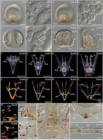
| Description | FIGURE 9. Skeletogenesis in Paracentrotus lividus during the embryonic and larval periods. Developmental stages are as follows: (A,B) early gastrula stage (EG); (C,D) mid-gastrula stage (mid-G); (E,F) late gastrula stage (LG); (G,H) prism stage (prism); (I,I') 2-arm pluteus stage (2-arm); (J,J′) 4-arm pluteus stage (4-arm); (K,K',M–P) 6-arm pluteus stage (6-arm); (L,L',Q–S) 8-arm pluteus stage (8-arm). In (A–L,N,P–S), images were acquired using light microscopy either in bright-field for (A–H,N,P–S), or dark-field for (I–L). In (I'–L',M,O), images were obtained using polarized light to highlight the skeletal elements. In (A,C,H), embryos are in right view, with the animal pole up and, for (H), with the ventral side left. In (E,G), embryos are in ventral view, with the animal pole up. In (I–L′), larvae are in anterior view, with the ventral side up. (B) Close-up of the skeletogenic mesoderm cells and their related filopodia in a ventral chain. (D,F) Close-ups of a ventrolateral cluster to highlight the rhombohedral crystal in (D) and the triradiate spicule in (F), the two images corresponding, respectively, to the regions highlighted by yellow boxes in (C) and (E). (M–P) Close-ups of the regions outlined by orange boxes in (K) with larvae in lateral view, except for (M) where the larva is in anterior view. (M,N) Close-ups of the developing posterodorsal spicule. (O,P) Close-ups of the developing dorsal arch. (Q–S) Close-ups of the regions highlighted by orange boxes in (L) with larvae in anterior view. (Q) Close-up of the skeletal elements located in the vicinity of the larval digestive tract. (R) Close-up of the body rods in the most dorsal region of the larva. (S) Close-up of the non-fenestrated spicule of an anterolateral arm. In (B), arrowheads highlight the filopodia extended by skeletogenic mesoderm cells. In (C–F), double arrowheads mark the skeletal elements forming in the ventrolateral clusters. In (H), the arrowhead highlights the limit between the right dorsoventral connecting rod and the right anterolateral rod. In (I,J), the asterisk marks the oral hood. Scale bar: (A,C,E,G,H,M–S) 30 μm; (B,D,F) 15 μm; (I) 100 μm; (J–L,J′–L′) 200 µm. AmR: anteromedial rod; Apx: apex; DA: dorsal arch; lAdtR: left anterodorsal transverse rod; lAla: left anterolateral arm; lAlR: left anterolateral rod; lAvtR: left anteroventral transverse rod; lBR: left body rod; lDvcR: left dorsoventral connecting rod; lPoa: left postoral arm; lPoR: left postoral rod; lPda: left posterodorsal arm; lPdR: left posterodorsal rod; lPra: left preoral arm; lPrR: left preoral rod; lRR: left recurrent rod; lVtR: left ventral transverse rod; rAdtR: right anterodorsal transverse rod; rAla: right anterolateral arm; rAlR: right anterolateral rod; rAvtR: right anteroventral transverse rod; rBR: right body rod; rDvcR: right dorsoventral connecting rod; rPoa: right postoral arm; rPoR: right postoral rod; rPda: right posterodorsal arm; rPdR: right posterodorsal rod; rPra: right preoral arm; rPrR: right preoral rod; rRR: right recurrent rod; rVLC: right ventrolateral cluster; rVtR: right ventral transverse rod; St: stomach. |
| Date | |
| Source |
https://www.frontiersin.org/articles/10.3389/fcell.2022.966408/full Developmental atlas of the indirect-developing sea urchin Paracentrotus lividus: From fertilization to juvenile stages, Front. Cell Dev. Biol., 31 October 2022 Sec. Morphogenesis and Patterning Volume 10 - 2022, https://doi.org/10.3389/fcell.2022.966408 |
| Author | Laurent Formery, Axel Wakefield, Maeva Gesson, Ludovic Toisoul, Guy Lhomond, Laurent Gilletta, Régis Lasbleiz, Michael Schubert, Jenifer C. Croce1 |
Licensing[edit]
This file is licensed under the Creative Commons Attribution 4.0 International license.
- You are free:
- to share – to copy, distribute and transmit the work
- to remix – to adapt the work
- Under the following conditions:
- attribution – You must give appropriate credit, provide a link to the license, and indicate if changes were made. You may do so in any reasonable manner, but not in any way that suggests the licensor endorses you or your use.

|
This file, which was originally posted to an external website, has not yet been reviewed by an administrator or reviewer to confirm that the above license is valid. See Category:License review needed for further instructions.
|
File history
Click on a date/time to view the file as it appeared at that time.
| Date/Time | Thumbnail | Dimensions | User | Comment | |
|---|---|---|---|---|---|
| current | 22:25, 5 March 2024 | | 1,243 × 1,675 (1.63 MB) | Rasbak (talk | contribs) | {{Information |description=FIGURE 9. Skeletogenesis in Paracentrotus lividus during the embryonic and larval periods. Developmental stages are as follows: (A,B) early gastrula stage (EG); (C,D) mid-gastrula stage (mid-G); (E,F) late gastrula stage (LG); (G,H) prism stage (prism); (I,I') 2-arm pluteus stage (2-arm); (J,J′) 4-arm pluteus stage (4-arm); (K,K',M–P) 6-arm pluteus stage (6-arm); (L,L',Q–S) 8-arm pluteus stage (8-arm). In (A–L,N,P–S), images were acquired using light microscopy eith... |
You cannot overwrite this file.
File usage on Commons
The following 2 pages use this file:
File usage on other wikis
The following other wikis use this file:
- Usage on nl.wikipedia.org