File:Perfused Rat Liver.JPG

Original file (2,448 × 1,632 pixels, file size: 399 KB, MIME type: image/jpeg)
Captions
Captions
From english wikipedia: http://en.wikipedia.org/wiki/Image:Perfused_Rat_Liver.JPG
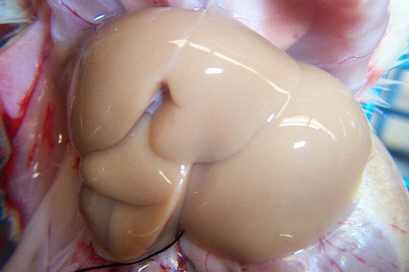
Photo of a Perfused Rat Liver
- You are free:
- to share – to copy, distribute and transmit the work
- to remix – to adapt the work
- Under the following conditions:
- attribution – You must give appropriate credit, provide a link to the license, and indicate if changes were made. You may do so in any reasonable manner, but not in any way that suggests the licensor endorses you or your use.
This photo was taken by CM North of Michigan State University during the isolation of hepatocytes from a rat liver using an Kodak DX4900 camera on 2004-04-27 and is usuable under the Creative Commons license. It appears beige because much of the blood has been cleared from circulation in preparation for cellular isolation. A normal liver has a red coloration to it. This is also significant glare from the fluorescent lights, but the image seems clear enough to
It should be noted that lobe configuration for a rat liver is also somewhat different from human, but visualization is still valuable for the sake of education. The liver is an organ in vertebrates including humans. It plays a major role in metabolism and has a number of functions in the body including detoxification, glycogen storage and plasma protein synthesis. It also produces bile which is very important for digestion. Medical terms related to the liver often start in hepato or hepatic from the greek word hepar for liver.
File history
Click on a date/time to view the file as it appeared at that time.
| Date/Time | Thumbnail | Dimensions | User | Comment | |
|---|---|---|---|---|---|
| current | 12:55, 7 November 2005 | | 2,448 × 1,632 (399 KB) | Snek01 (talk | contribs) | Photo of a Perfused Rat Liver{{cc-by}} This photo was taken by CM North of Michigan State University during the isolation of hepatocytes from a rat liver using an Kodak DX4900 camera on 2004-04-27 and is usuable under the Creative Commons license. It app |
You cannot overwrite this file.
File usage on Commons
There are no pages that use this file.
File usage on other wikis
The following other wikis use this file: