File:Photomicrographs of calbindin and parvalbumin immunoreactive neurons in the nucleus accumbens.JPEG
Jump to navigation
Jump to search

Size of this preview: 423 × 600 pixels. Other resolutions: 169 × 240 pixels | 338 × 480 pixels | 542 × 768 pixels | 722 × 1,024 pixels | 1,444 × 2,048 pixels | 3,203 × 4,541 pixels.
Original file (3,203 × 4,541 pixels, file size: 1.5 MB, MIME type: image/jpeg)
Captions
Captions
Add a one-line explanation of what this file represents
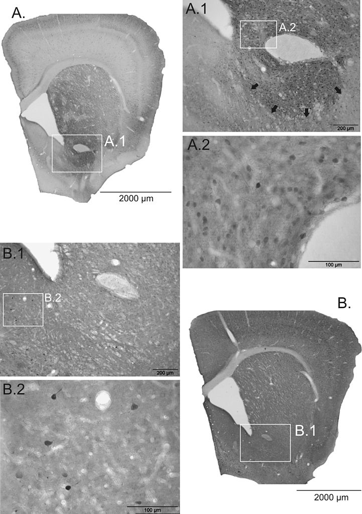
| Description | Comparative image to give an idea of calbindin and parvalbumin distribution in the nucleus accumbens. Figure legend in source article (CC-by-2.0): "Photomicrographs of Calbindin and Parvalbumin immunoreactive neurons in the nucleus accumbens. Overview (A) and higher magnifications (A1, A2) of the Calbindin innervation of the NAC. The majority of CB+ cells is located in the core, which border to the shell is detectable (black arrows). PV+ cells are almost exclusively located in the shell (B1, B2), however, the overall density is much lower compared to CB+ cells. Scale bars: 2000 μm (A, B); 200 μm (A1, B1); 100 μm (A2, B2)." |
| Date | |
| Source | Brummelte, Susanne; Thorsten Grund, Andrea Czok, Gertraud Teuchert-Noodt, Jorg Neddens (2006). "Long-term effects of a single adult methamphetamine challenge: Minor impact on dopamine fibre density in limbic brain areas of gerbils". Behavioral and Brain Functions 2 (1): 12. DOI:10.1186/1744-9081-2-12. ISSN 1744-9081. Retrieved on 2007-12-21. |
| Author | Article by Susanne Brummelte, Thorsten Grund, Andrea Czok, Gertraud Teuchert-Noodt, Jorg Neddens |
| Permission (Reusing this file) |
This file is licensed under the Creative Commons Attribution 2.0 Generic license.
|
File history
Click on a date/time to view the file as it appeared at that time.
| Date/Time | Thumbnail | Dimensions | User | Comment | |
|---|---|---|---|---|---|
| current | 21:52, 21 December 2007 | | 3,203 × 4,541 (1.5 MB) | OldakQuill (talk | contribs) | {{Information |Description=Comparative image to give an idea of calbindin and parvalbumin distribution in the nucleus accumbens. Figure legend in source article (CC-by-2.0): "'''Photomicrographs of Calbindin and Parvalbumin immunoreactive neurons in the n |
You cannot overwrite this file.
File usage on Commons
There are no pages that use this file.