File:Picomonas judraskeda.png
Jump to navigation
Jump to search
Size of this preview: 440 × 599 pixels. Other resolutions: 176 × 240 pixels | 352 × 480 pixels | 564 × 768 pixels | 752 × 1,024 pixels | 1,504 × 2,048 pixels | 3,098 × 4,217 pixels.
Original file (3,098 × 4,217 pixels, file size: 9.71 MB, MIME type: image/png)
Captions
Captions
Add a one-line explanation of what this file represents
Summary[edit]
| Description |
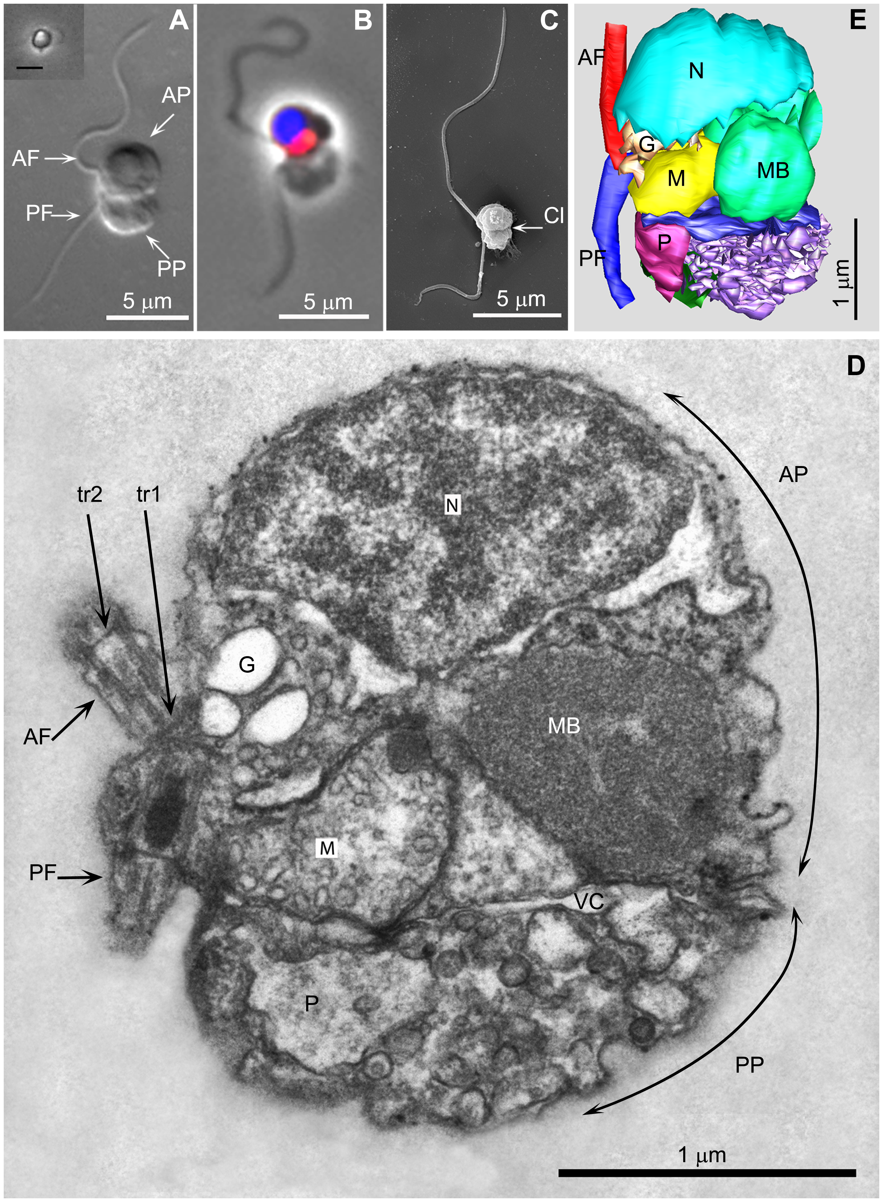
English: A Picomonas cell.
|
||
| Date | |||
| Source | doi:10.1371/journal.pone.0059565 | ||
| Author | Ramkumar Seenivasan, Nicole Sausen, Linda K. Medlin, Michael Melkonian | ||
| Permission (Reusing this file) |
|
File history
Click on a date/time to view the file as it appeared at that time.
| Date/Time | Thumbnail | Dimensions | User | Comment | |
|---|---|---|---|---|---|
| current | 09:17, 29 August 2013 | | 3,098 × 4,217 (9.71 MB) | Alexander Shatulin (talk | contribs) | Higher resolution |
| 12:14, 16 April 2013 |  | 441 × 600 (314 KB) | Haplochromis (talk | contribs) | {{Information |Description ={{en|1=A ''Picomonas'' cell. 2A. Differential interference contrast of a chemically fixed cell. Inset shows phase contrast image of a live cell from tissue culture flask photographed with an inverted microscope (Scale ba... |
You cannot overwrite this file.
File usage on Commons
The following 2 pages use this file:
File usage on other wikis
The following other wikis use this file:
- Usage on bg.wikipedia.org
- Usage on bs.wikipedia.org
- Usage on cs.wikipedia.org
- Usage on de.wikipedia.org
- Usage on es.wikipedia.org
- Usage on he.wikipedia.org
- Usage on ia.wikipedia.org
- Usage on ko.wikipedia.org
- Usage on ru.wikipedia.org
- Usage on www.wikidata.org