File:Pseudoachondroplasia. 02.jpg
Jump to navigation
Jump to search
Size of this preview: 500 × 600 pixels. Other resolutions: 200 × 240 pixels | 400 × 480 pixels | 640 × 768 pixels | 853 × 1,024 pixels | 1,600 × 1,920 pixels.
Original file (1,600 × 1,920 pixels, file size: 171 KB, MIME type: image/jpeg)
Captions
Captions
Add a one-line explanation of what this file represents
More images of this case:  
|
Summary
[edit]
| Description |
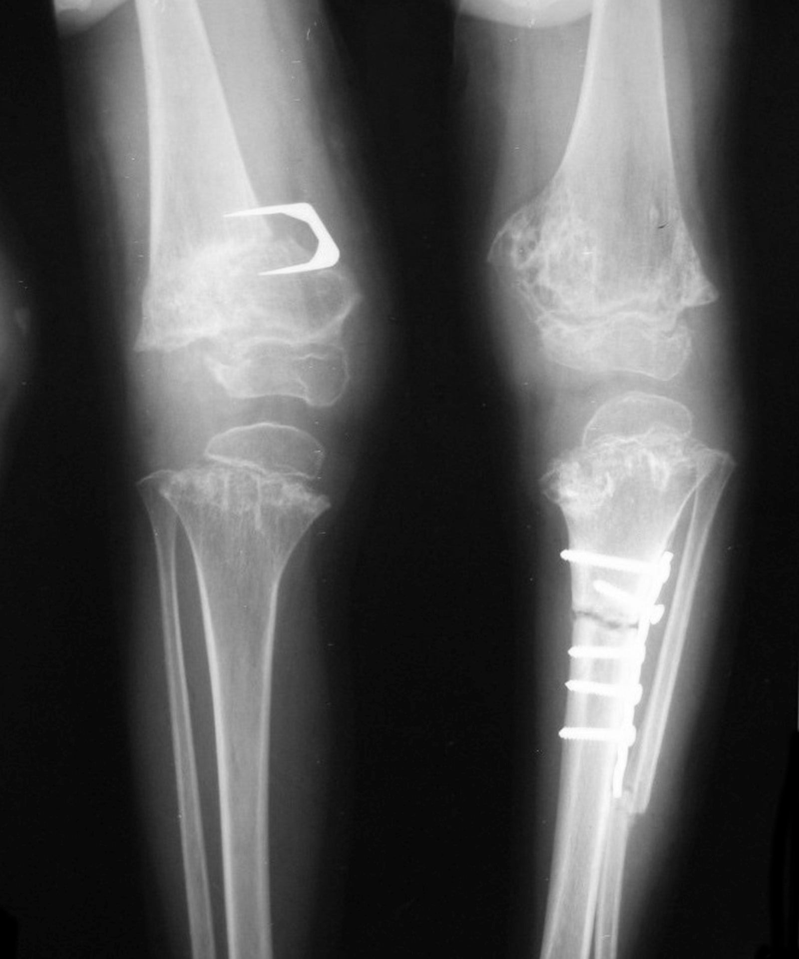
English: Pseudoachondroplasia. Knee radiographs AP view of a child depicting dysplastic distal femoral and proximal tibial epiphyses, with distal femoral metaphyseal broadening and irregularities. Note the metaphyseal lines of ossification of the proximal tibias and distal femora and relative sparing of the tibial shafts. The changes around the knee are known as "rachitic-like changes". Lesions are fairly symmetrical. Note the internal fixation implants used to correct a femoral valgus and tibial varus deformity. |
| Date | |
| Source | Own work |
| Author | Bonejoint |
Licensing
[edit]
I, the copyright holder of this work, hereby publish it under the following license:
This file is licensed under the Creative Commons Attribution-Share Alike 4.0 International license.
- You are free:
- to share – to copy, distribute and transmit the work
- to remix – to adapt the work
- Under the following conditions:
- attribution – You must give appropriate credit, provide a link to the license, and indicate if changes were made. You may do so in any reasonable manner, but not in any way that suggests the licensor endorses you or your use.
- share alike – If you remix, transform, or build upon the material, you must distribute your contributions under the same or compatible license as the original.
File history
Click on a date/time to view the file as it appeared at that time.
| Date/Time | Thumbnail | Dimensions | User | Comment | |
|---|---|---|---|---|---|
| current | 20:33, 18 September 2021 | | 1,600 × 1,920 (171 KB) | Hellerhoff (talk | contribs) | color not needed, small retouche |
| 19:46, 10 October 2017 |  | 1,600 × 1,920 (262 KB) | Bonejoint (talk | contribs) | This file shows radiologic abnormalities of of pseudoachondroplasia in a child together with internal fixation of the corrective osteotomies that address the underlying knee deformities. | |
| 19:04, 19 April 2017 |  | 3,728 × 3,894 (471 KB) | Bonejoint (talk | contribs) | User created page with UploadWizard |
You cannot overwrite this file.
File usage on Commons
The following 2 pages use this file:
File usage on other wikis
The following other wikis use this file:
- Usage on en.wikipedia.org
- Usage on mk.wikipedia.org