File:Rabbit uterus during pseudopregnancy showing, telocytes.png
Jump to navigation
Jump to search

Size of this preview: 800 × 395 pixels. Other resolutions: 320 × 158 pixels | 640 × 316 pixels | 1,376 × 679 pixels.
Original file (1,376 × 679 pixels, file size: 766 KB, MIME type: image/png)
Captions
Captions
Add a one-line explanation of what this file represents
Summary
[edit]
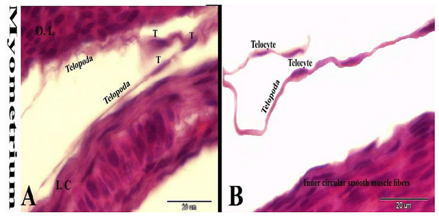
| Description | Figure 7. Photomicrograph of the rabbit uterus during pseudopregnancy showing, telocytes (T) with its characteristic telopoda in the myometrium between the inner circular and outer longitudinal smooth muscle fibers of the myometrium. Original magnification, A & B: 1000X, scale bar=20 µm, Hematoxylin and Eosin stain. |
| Date | |
| Source | https://www.researchgate.net/publication/316631217_Morphological_Histological_and_Immunohistochemical_Study_of_the_Rabbit_Uterus_during_Pseudopregnancy Morphological, Histological and Immunohistochemical Study of the Rabbit Uterus during Pseudopregnancy. J Cytol Histol 8: 443. doi: 10.4172/2157-7099.1000443 |
| Author | Abd-Elkareem MD |

|
This file, which was originally posted to an external website, has not yet been reviewed by an administrator or reviewer to confirm that the above license is valid. See Category:License review needed for further instructions.
|
© 2017 Abd-Elkareem MD. This is an open-access article distributed under the terms of the Creative Commons Attribution License, which permits unrestricted use, distribution, and reproduction in any medium, provided the original author and source are credited.
Licensing
[edit]
This file is licensed under the Creative Commons Attribution 4.0 International license.
- You are free:
- to share – to copy, distribute and transmit the work
- to remix – to adapt the work
- Under the following conditions:
- attribution – You must give appropriate credit, provide a link to the license, and indicate if changes were made. You may do so in any reasonable manner, but not in any way that suggests the licensor endorses you or your use.
File history
Click on a date/time to view the file as it appeared at that time.
| Date/Time | Thumbnail | Dimensions | User | Comment | |
|---|---|---|---|---|---|
| current | 20:56, 19 June 2024 | | 1,376 × 679 (766 KB) | Rasbak (talk | contribs) | {{Information |description=Figure 7. Photomicrograph of the rabbit uterus during pseudopregnancy showing, telocytes (T) with its characteristic telopoda in the myometrium between the inner circular and outer longitudinal smooth muscle fibers of the myometrium. Original magnification, A & B: 1000X, scale bar=20 µm, Hematoxylin and Eosin stain. |date=2017-03-15 |source=https://www.researchgate.net/publication/316631217_Morphological_Histological_and_Immunohistochemical_Study_of_the_Rabbit_Uter... |
You cannot overwrite this file.
File usage on Commons
There are no pages that use this file.
File usage on other wikis
The following other wikis use this file:
- Usage on nl.wikipedia.org