File:Immunohistology of the subfornical organ in a healthy SJL mouse.jpg
(Redirected from File:Sfo.jpg)

Size of this preview: 215 × 598 pixels. Other resolutions: 86 × 240 pixels | 172 × 480 pixels | 709 × 1,971 pixels.
Original file (709 × 1,971 pixels, file size: 476 KB, MIME type: image/jpeg)
Captions
Captions
Add a one-line explanation of what this file represents
| Description |
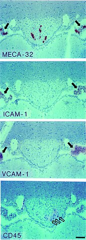
English: Immunohistology of the en:subfornical organ (SFO) in the healthy SJL mouse. The top panel shows MECA-32+ capillaries within the SFO. Panels below demonstrate that ICAM-1 and VCAM-1 can not be detected on the MECA-32+ capillaries within the SFO. Single CD45+ perivascular cells can be detected within the SFO (open arrows, lower panel). Note the positive immunostaining for MECA-32 on en:choroid plexus capillaires and for ICAM-1 and VCAM-1 on the choroid plexus epithelium visible at the left and right margins (closed arrows). Immunoperoxidase, hematoxylin counterstain. Bar = 100 μm. This result was observed 10 times.
Polski: Immunohistologia narządu podspoidłowego (SFO) u myszy SJL. Pierwszy panel pokazuje reakcję naczyń SFO z przeciwciałami anty-MECA-32, kolejne dwa panele obrazują dodatnią reakcję na obecność ICAM-1 i VCAM-1. W dolnym panelu pojedyczne komórki są CD45+ (białe strzałki). Po bokach od SFO widać we wszystkich preparatach dodatnie reakcje komórek splotu naczyniówkowego (ciemne strzałki). Podziałka wynosi 100 μm. |
| Date | 3 May 2008 (upload date) |
| Source | Schulz M, Engelhardt B. The circumventricular organs participate in the immunopathogenesis of experimental autoimmune encephalomyelitis. Cerebrospinal Fluid Res. 2, 8. 2005. doi:10.1186/1743-8454-2-8. PMID 16197544 |
| Author | Schulz M, Engelhardt B. |
| Permission (Reusing this file) |
This file is licensed under the Creative Commons Attribution 2.0 Generic license.
|
File history
Click on a date/time to view the file as it appeared at that time.
| Date/Time | Thumbnail | Dimensions | User | Comment | |
|---|---|---|---|---|---|
| current | 12:16, 3 May 2008 | 709 × 1,971 (476 KB) | Filip em (talk | contribs) | {{Information |Description=Immunohistology of the subfornical organ (SFO) in the healthy SJL mouse. The top panel shows MECA-32+ capillaries within the SFO. Panels below demonstrate that ICAM-1 and VCAM-1 can not be detected on the MECA-32+ capillaries wi |
You cannot overwrite this file.
File usage on Commons
The following page uses this file:
- File:Sfo.jpg (file redirect)
File usage on other wikis
The following other wikis use this file:
- Usage on pl.wikipedia.org