File:Sheep midbrain.jpg
Jump to navigation
Jump to search

Size of this preview: 669 × 599 pixels. Other resolutions: 268 × 240 pixels | 536 × 480 pixels | 857 × 768 pixels | 1,200 × 1,075 pixels.
Original file (1,200 × 1,075 pixels, file size: 334 KB, MIME type: image/jpeg)
Captions
Captions
Add a one-line explanation of what this file represents
| Description |
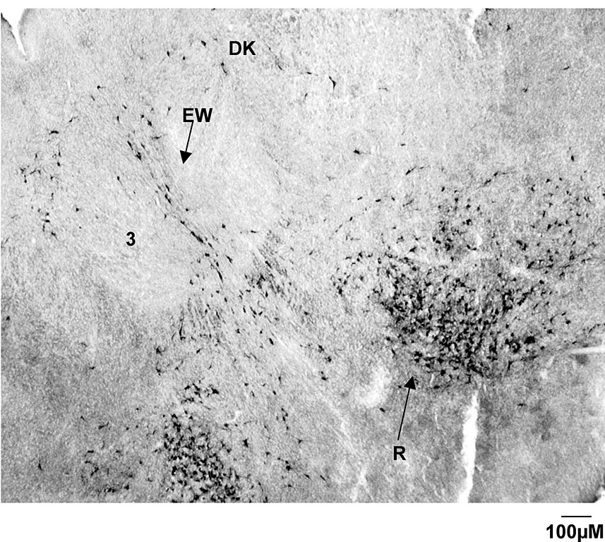
English: CTB labelling in the red nucleus of a G55 fetus. Note the CTB labelling is bilateral, and forms a distinct tight cluster, with a greater density of cells towards the ventral region of the nucleus. Labelling is also observed in the nucleus of Darkschewitsch (Dk) and in the vicinity of the Edinger-Westphal nucleus (EW). 3 = oculomotor nucleus. |
| Date | |
| Source | Stockx EM, Anderson CR, Murphy SM, Cooke IR, Berger PJ. The development of descending projections from the brainstem to the spinal cord in the fetal sheep. BMC neuroscience: 40 (styczeń 2007). doi:10.1186/1471-2202-8-40. PMID 17577416 |
| Author | see below |
| Permission (Reusing this file) |
[1] |
This file is licensed under the Creative Commons Attribution 2.0 Generic license.
- You are free:
- to share – to copy, distribute and transmit the work
- to remix – to adapt the work
- Under the following conditions:
- attribution – You must give appropriate credit, provide a link to the license, and indicate if changes were made. You may do so in any reasonable manner, but not in any way that suggests the licensor endorses you or your use.
File history
Click on a date/time to view the file as it appeared at that time.
| Date/Time | Thumbnail | Dimensions | User | Comment | |
|---|---|---|---|---|---|
| current | 21:06, 11 January 2009 | | 1,200 × 1,075 (334 KB) | Filip em (talk | contribs) | {{Information |Description={{en|1=CTB labelling in the red nucleus of a G55 fetus. Note the CTB labelling is bilateral, and forms a distinct tight cluster, with a greater density of cells towards the ventral region of the nucleus. Labelling is also observ |
You cannot overwrite this file.
File usage on Commons
There are no pages that use this file.
File usage on other wikis
The following other wikis use this file:
- Usage on pl.wikipedia.org