File:Squamous Ovarian Cell Carcinoma.png
Jump to navigation
Jump to search
Size of this preview: 800 × 463 pixels. Other resolutions: 320 × 185 pixels | 640 × 370 pixels | 1,024 × 593 pixels | 1,280 × 741 pixels | 2,560 × 1,481 pixels | 3,776 × 2,185 pixels.
Original file (3,776 × 2,185 pixels, file size: 8.18 MB, MIME type: image/png)
Captions
Captions
Add a one-line explanation of what this file represents
Summary
[edit]
| Description |
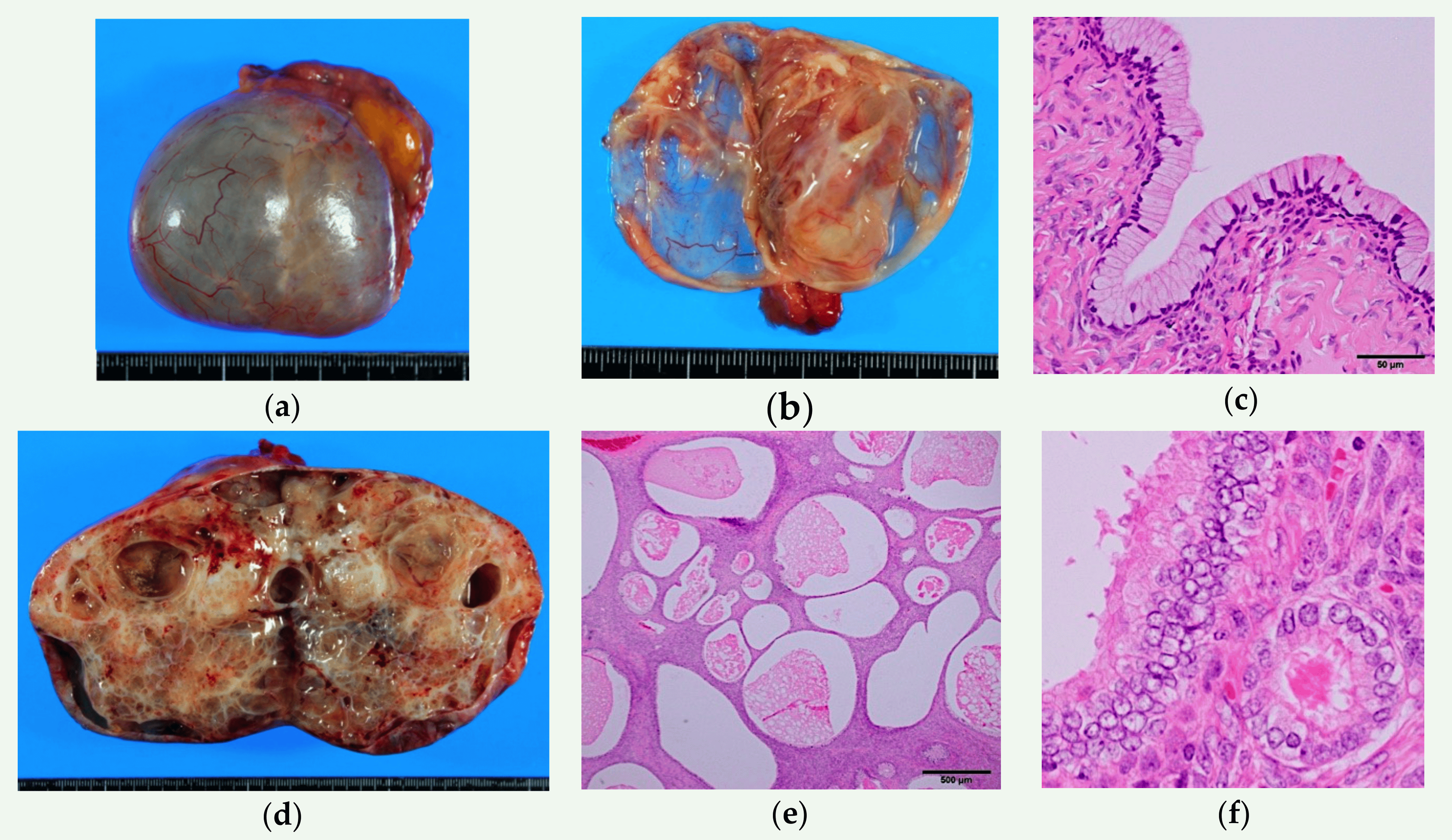
English: Gross and microscopic appearance of benign seromucous tumors. (a–c) Seromucinous cystadenoma. A thin-walled paucilocular cyst filled with thin mucinous clear fluid. The cyst wall is lined by endocervical-type columnar mucinous cells admixed with scattered ciliated cells. Note the resemblance to normal endocervical epithelium (d–f) Seromucinous adenofibroma. Mostly solid mass with scattered cystic spaces. Endocervical-type cells proliferate in an adenofibromatous pattern. Focal nuclear enlargement and stratifications—but no papillary projections—are seen. |
| Date | |
| Source | Nagamine, M.; Mikami, Y. Ovarian Seromucinous Tumors: Pathogenesis, Morphologic Spectrum, and Clinical Issues. Diagnostics 2020, 10, 77. https://doi.org/10.3390/diagnostics10020077 |
| Author | Nagamine, M.; Mikami, Y |
Licensing
[edit]
This file is licensed under the Creative Commons Attribution 4.0 International license.
- You are free:
- to share – to copy, distribute and transmit the work
- to remix – to adapt the work
- Under the following conditions:
- attribution – You must give appropriate credit, provide a link to the license, and indicate if changes were made. You may do so in any reasonable manner, but not in any way that suggests the licensor endorses you or your use.

|
This media file is uncategorized.
Please help improve this media file by adding it to one or more categories, so it may be associated with related media files (how?), and so that it can be more easily found.
Please notify the uploader with {{subst:Please link images|File:Squamous Ovarian Cell Carcinoma.png}} ~~~~ |
File history
Click on a date/time to view the file as it appeared at that time.
| Date/Time | Thumbnail | Dimensions | User | Comment | |
|---|---|---|---|---|---|
| current | 18:25, 20 November 2023 | | 3,776 × 2,185 (8.18 MB) | Cheyleann (talk | contribs) | Uploaded a work by Nagamine, M.; Mikami, Y from Nagamine, M.; Mikami, Y. Ovarian Seromucinous Tumors: Pathogenesis, Morphologic Spectrum, and Clinical Issues. Diagnostics 2020, 10, 77. https://doi.org/10.3390/diagnostics10020077 with UploadWizard |
You cannot overwrite this file.
File usage on Commons
There are no pages that use this file.
File usage on other wikis
The following other wikis use this file:
- Usage on en.wikipedia.org