File:Umbilical Granulation.jpg

Original file (484 × 621 pixels, file size: 241 KB, MIME type: image/jpeg)
Captions
Captions
Summary
[edit]
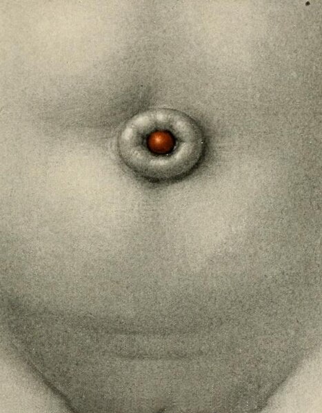
| Description | Fig. 65. — An Umbilical Granulation. The umbilical ring is unusually prominent, protruding at least 2 mm. above the abdominal wall. In the center is a small, globular, red mass. It was very friable, was readily removed, and did not recur. On histologic examination it was found to consist essentially of young granulation tissue rich in blood-capillaries. It contained no epithelial elements. |
| Date | |
| Source | https://archive.org/details/embryologyanatom00cull/page/116/mode/2up Embryology, anatomy, and diseases of the umbilicus : together with diseases of the urachus |
| Author | Cullen, Thomas Stephen, 1868-1953 |
Licensing
[edit]
|
This work is in the public domain in its country of origin and other countries and areas where the copyright term is the author's life plus 70 years or fewer.
| |
| This file has been identified as being free of known restrictions under copyright law, including all related and neighboring rights. | |

|
This file, which was originally posted to an external website, has not yet been reviewed by an administrator or reviewer to confirm that the above license is valid. See Category:License review needed for further instructions.
|
File history
Click on a date/time to view the file as it appeared at that time.
| Date/Time | Thumbnail | Dimensions | User | Comment | |
|---|---|---|---|---|---|
| current | 18:28, 18 February 2024 | | 484 × 621 (241 KB) | Rasbak (talk | contribs) | {{Information |description=Fig. 65. — An Umbilical Granulation. The umbilical ring is unusually prominent, protruding at least 2 mm. above the abdominal wall. In the center is a small, globular, red mass. It was very friable, was readily removed, and did not recur. On histologic examination it was found to consist essentially of young granulation tissue rich in blood-capillaries. It contained no epithelial elements. |date= 1916 |source=https://archive.org/details/embryologyanatom00cull/page/1... |
You cannot overwrite this file.
File usage on Commons
There are no pages that use this file.
File usage on other wikis
The following other wikis use this file:
- Usage on nl.wikipedia.org