File:Chlamydomonas TEM 09.jpg
Jump to navigation
Jump to search

Size of this preview: 751 × 600 pixels. Other resolutions: 301 × 240 pixels | 601 × 480 pixels | 961 × 768 pixels | 1,280 × 1,023 pixels | 1,800 × 1,438 pixels.
Original file (1,800 × 1,438 pixels, file size: 784 KB, MIME type: image/jpeg)
Captions
Captions
Add a one-line explanation of what this file represents
| Description |
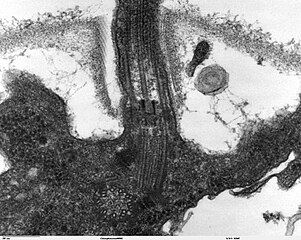
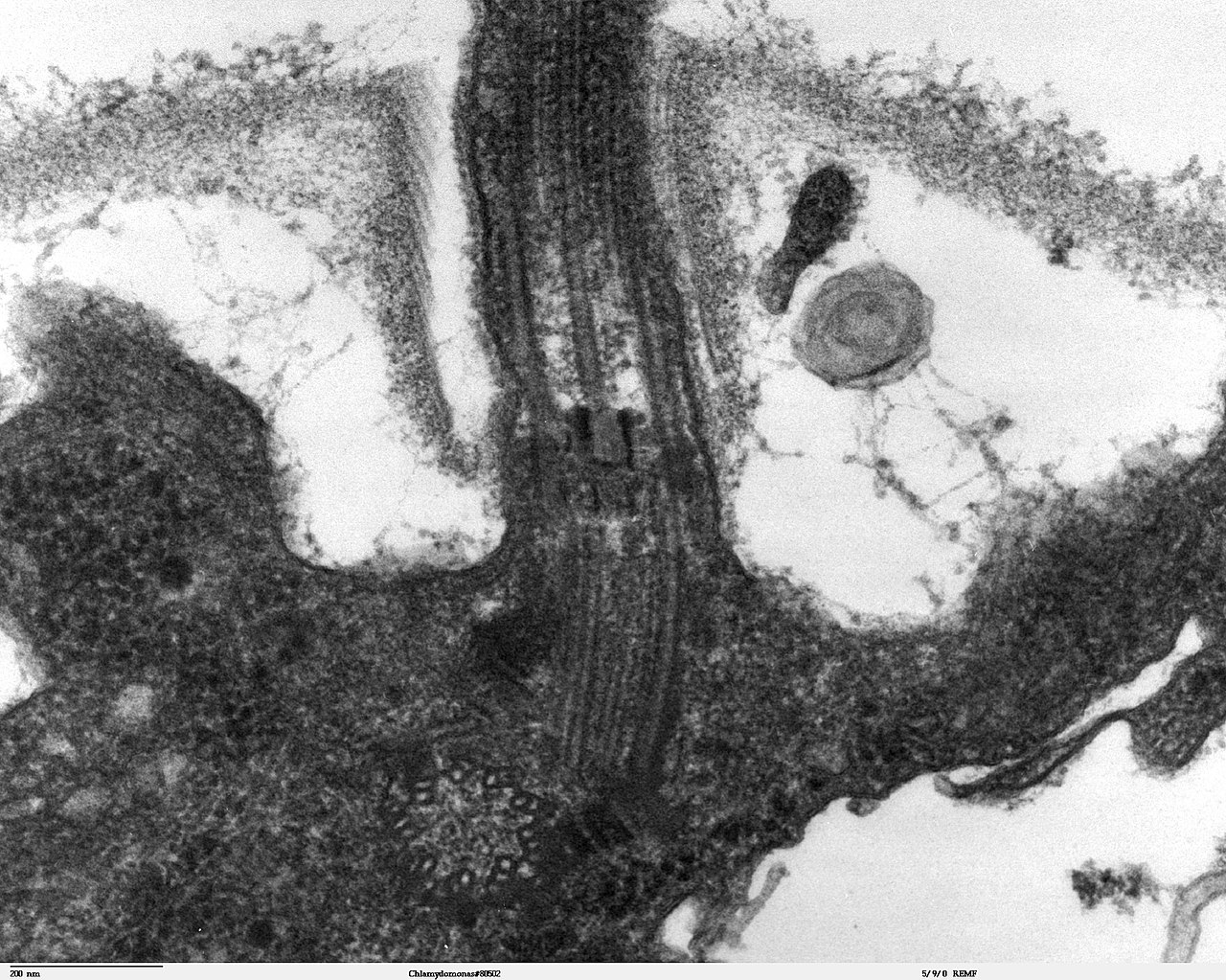
Transmission electron microscope image, showing an example of green algae (Chlorophyta). Chlamydomanas reinhardtii is a unicellular flagellate used as a model system in molecular genetics work and flagellar motility studies. This image is a longitudinal section through the flagella area. In the cell apex is the basal body that is the anchoring site for a flagella. Basal bodies originate from and have a substructure similar to that of centrioles, with nine peripheral microtubule triplets(see structure at bottom center of image). The two inner microtubules of each triplet in a basal body become the two outer doublets in the flagella. This image also shows the transition region, with its fibers of the stellate structure. The top of the image shows the flagella passing through the cell wall. |
| Date | |
| Source | Source and public domain notice at http://remf.dartmouth.edu/imagesindex.html |
| Author | Dartmouth Electron Microscope Facility, Dartmouth College |
| Permission (Reusing this file) |
Released into the public domain |
| This work has been released into the public domain by its author, Dartmouth Electron Microscope Facility, Dartmouth College. This applies worldwide. In some countries this may not be legally possible; if so: Dartmouth Electron Microscope Facility, Dartmouth College grants anyone the right to use this work for any purpose, without any conditions, unless such conditions are required by law.
|
File history
Click on a date/time to view the file as it appeared at that time.
| Date/Time | Thumbnail | Dimensions | User | Comment | |
|---|---|---|---|---|---|
| current | 06:47, 21 September 2007 | | 1,800 × 1,438 (784 KB) | Neil916 (talk | contribs) | {{Information |Description= Transmission electron microscope image, showing an example of green algae (Chlorophyta). <br><br>''Chlamydomanas reinhardtii'' is a unicellular flagellate used as a model system in molecular genetics work and flagellar motilit |
You cannot overwrite this file.
File usage on Commons
There are no pages that use this file.
File usage on other wikis
The following other wikis use this file:
- Usage on ar.wikipedia.org
- Usage on bs.wikipedia.org
- Usage on ca.wikipedia.org
- Usage on cs.wikipedia.org
- Usage on de.wikipedia.org
- Usage on de.wikibooks.org
- Usage on en.wikipedia.org
- Usage on es.wikipedia.org
- Usage on gl.wikipedia.org
- Usage on id.wikipedia.org
- Usage on ja.wikipedia.org
- Usage on ko.wikipedia.org
- Usage on pl.wikipedia.org
- Usage on ru.wikipedia.org
- Usage on sv.wikipedia.org
- Usage on tr.wikipedia.org
- Usage on uk.wikipedia.org
- Usage on zh.wikipedia.org